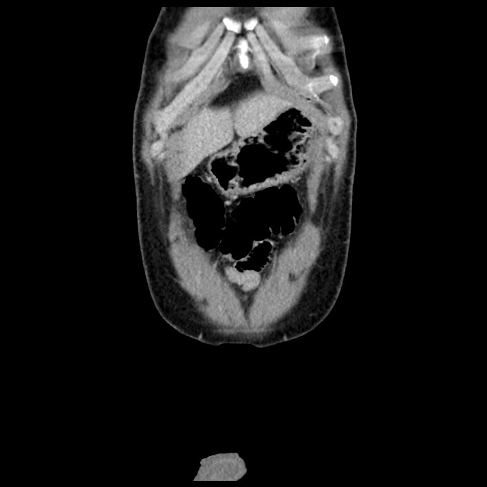
File:Co-existing acute appendicitis and epiploic appendagitis (Radiopaedia 61789-69911 C 7).jpg
Jump to navigation
Jump to search
Size of this preview: 600 × 600 pixels. Other resolutions: 240 × 240 pixels | 480 × 480 pixels | 708 × 708 pixels.
Original file (708 × 708 pixels, file size: 41 KB, MIME type: image/jpeg)
Summary:
| Description |
|
| Date | Published: 7th Aug 2018 |
| Source | https://radiopaedia.org/cases/co-existing-acute-appendicitis-and-epiploic-appendagitis |
| Author | Ahmed Samir |
| Permission (Permission-reusing-text) |
http://creativecommons.org/licenses/by-nc-sa/3.0/ |
Licensing:
Attribution-NonCommercial-ShareAlike 3.0 Unported (CC BY-NC-SA 3.0)
File history
Click on a date/time to view the file as it appeared at that time.
| Date/Time | Thumbnail | Dimensions | User | Comment | |
|---|---|---|---|---|---|
| current | 00:08, 30 August 2021 | | 708 × 708 (41 KB) | Fæ (talk | contribs) | Radiopaedia project rID:61789 (batch #8564-190 C7) |
You cannot overwrite this file.
File usage
The following page uses this file:
.jpg&oldid=1187064)