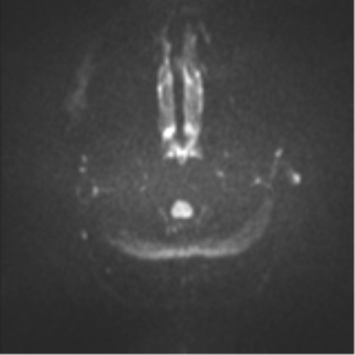
File:Colloid cyst (Radiopaedia 44510-48181 Axial DWI 29).png
Jump to navigation
Jump to search
Colloid_cyst_(Radiopaedia_44510-48181_Axial_DWI_29).png (512 × 512 pixels, file size: 139 KB, MIME type: image/png)
Summary:
| Description |
|
| Date | Published: 29th Sep 2016 |
| Source | https://radiopaedia.org/cases/colloid-cyst-20 |
| Author | Frank Gaillard |
| Permission (Permission-reusing-text) |
http://creativecommons.org/licenses/by-nc-sa/3.0/ |
Licensing:
Attribution-NonCommercial-ShareAlike 3.0 Unported (CC BY-NC-SA 3.0)
File history
Click on a date/time to view the file as it appeared at that time.
| Date/Time | Thumbnail | Dimensions | User | Comment | |
|---|---|---|---|---|---|
| current | 12:42, 30 August 2021 |  | 512 × 512 (139 KB) | Fæ (talk | contribs) | Radiopaedia project rID:44510 (batch #8630-135 E29) |
You cannot overwrite this file.
File usage
The following page uses this file:
