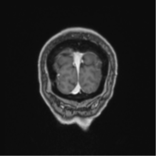
File:Colloid cyst of the third ventricle (Radiopaedia 86571-102662 Coronal T1 C+ 9).png

From NC Commons
Jump to navigation Jump to search

Colloid_cyst_of_the_third_ventricle_(Radiopaedia_86571-102662_Coronal_T1_C+_9).png(511 × 512 pixels, file size: 119 KB, MIME type: image/png)

Summary:

Description
  • Radiopaedia case ID: 86571
  • Image ID: 54340593
  • Image stack position: 9/90
  • Plane projection: Coronal
  • Aux modality: T1 C+
  • Study findings: 12 x 10 x 13mm lesion located at the roof of the foramen of Munro. This is isointense on T1-weighted sequence and hyperintense to T2-weighted sequence relative to the white matter.  There is enlarged lateral ventricle consistent with obstructive hydrocephalus. No transependymal edema. Grey-white matter differentiation is preserved. No abnormal restricted diffusion or susceptibility. Conclusion: 13mm colloid cyst at the foramen of Munro with associated obstructive hydrocephalus.
  • Modality: MRI
  • System: Central Nervous System
  • Findings: There is a hyperdense lesion centered at the foramen of Munro resulting in acute hydrocephalus with enlarged lateral ventricles and associated sulcal effacement. No transependymal edema. Cerebral aqueduct and the fourth ventricle remain patent. There is preservation of the grey-white matter differentiation. No midline shift. Conclusion: Colloid cyst of the third ventricle with associated obstructive hydrocephalus.
Date Published: 5th Feb 2021
Source https://radiopaedia.org/cases/colloid-cyst-of-the-third-ventricle-10
Author Kosuke Kato
Permission
(Permission-reusing-text)
http://creativecommons.org/licenses/by-nc-sa/3.0/

Licensing:

Attribution-NonCommercial-ShareAlike 3.0 Unported (CC BY-NC-SA 3.0)

File history

Click on a date/time to view the file as it appeared at that time.

Date/TimeThumbnailDimensionsUserComment
current01:24, 31 August 2021Thumbnail for version as of 01:24, 31 August 2021511 × 512 (119 KB) (talk | contribs)Radiopaedia project rID:86571 (batch #8644-534 J9)

Metadata