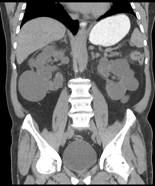
File:Colon adenocarcinoma - splenic flexure (Radiopaedia 34143-35406 B 40).png

From NC Commons
Jump to navigation Jump to search

Original file(512 × 615 pixels, file size: 304 KB, MIME type: image/png)

Summary:

Description
  • Radiopaedia case ID: 34143
  • Image ID: 10910357
  • Image stack position: 40/66
  • Plane projection: Coronal
  • Aux modality: Oral contrast
  • Modality: CT
  • System: Gastrointestinal
  • Findings: CT Abdomen / Pelvis Technique: Volume acquisition through the abdomen and pelvis has been obtained with oral contrast only due to poor renal function. Findings: At the level of the splenic flexure, there is a 4 cm segment of circumferentially thickened colon, with a single focus of mural calcification. Immediately upstream of this thickening, the large bowel is mildly distended up to 6 cm. Distal to this, caliber of the remaining colon is normal. Surrounding this thickening, there is associated stranding of the pericolic fat. No intraabdominal lymphadenopathy. The small bowel has an unremarkable appearance with no evidence of small bowel distension. There are only scattered sigmoid diverticula, with no evidence of diverticulitis. There are innumerable bilateral renal cysts, almost all of them demonstrating simple water attenuation. Within the right kidney, there are 8 mm and 14 mm rounded hyperdensities that do not however demonstrate fluid attenuation. Within segment 3 of the liver, immediately adjacent to the falciform ligament, there is a 14 mm rounded hypodensity that demonstrates water attenuation. There is also a smaller subcentimeter cyst within segment 2. There is a 16 mm region of irregular hypodensity within the posterior aspect of segment 3. Tiny calcified granuloma noted within segment 6 of the liver. Possible subcentimeter stone within the neck of the gallbladder. The common bile duct is not dilated. The pancreas, spleen and adrenal glands have an unremarkable appearance on non-contrast imaging. No suspicious bony lesion. Conclusion: Circumferential mural thickening of the splenic flexure with surrounding fat stranding, is concerning for a colonic tumor. Mild upstream distension of the colon, suggests this is at least partially obstructing. Endoscopic evaluation should be considered. Bilateral polycystic kidneys. Two hyperdense lesions within the right kidney likely represent hemorrhagic cysts.
Date Published: 6th Feb 2015
Source https://radiopaedia.org/cases/colon-adenocarcinoma-splenic-flexure-1
Author Kenny Sim
Permission
(Permission-reusing-text)
http://creativecommons.org/licenses/by-nc-sa/3.0/

Licensing:

Attribution-NonCommercial-ShareAlike 3.0 Unported (CC BY-NC-SA 3.0)

File history

Click on a date/time to view the file as it appeared at that time.

Date/TimeThumbnailDimensionsUserComment
current00:36, 1 September 2021Thumbnail for version as of 00:36, 1 September 2021512 × 615 (304 KB) (talk | contribs)Radiopaedia project rID:34143 (batch #8692-134 B40)

Metadata