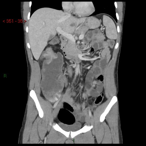
File:Colon cancer (Radiopaedia 56256-62927 B 34).jpg
Jump to navigation
Jump to search
Colon_cancer_(Radiopaedia_56256-62927_B_34).jpg (512 × 512 pixels, file size: 43 KB, MIME type: image/jpeg)
Summary:
| Description |
|
| Date | Published: 2nd Nov 2017 |
| Source | https://radiopaedia.org/cases/colon-cancer-1 |
| Author | Safwat Mohammad Almoghazy |
| Permission (Permission-reusing-text) |
http://creativecommons.org/licenses/by-nc-sa/3.0/ |
Licensing:
Attribution-NonCommercial-ShareAlike 3.0 Unported (CC BY-NC-SA 3.0)
File history
Click on a date/time to view the file as it appeared at that time.
| Date/Time | Thumbnail | Dimensions | User | Comment | |
|---|---|---|---|---|---|
| current | 07:26, 1 September 2021 |  | 512 × 512 (43 KB) | Fæ (talk | contribs) | Radiopaedia project rID:56256 (batch #8700-156 B34) |
You cannot overwrite this file.
File usage
The following page uses this file:
