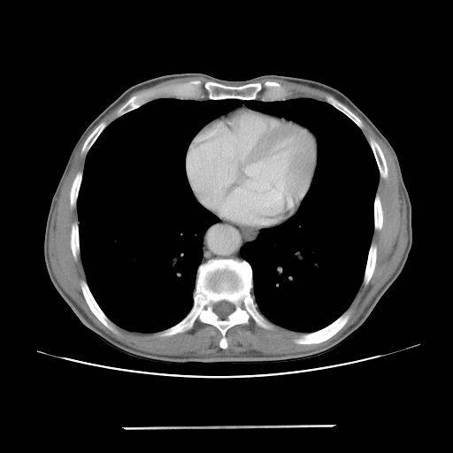
File:Colon cancer with calcified liver metastasis (Radiopaedia 74423-85307 A 3).jpg
Jump to navigation
Jump to search
Colon_cancer_with_calcified_liver_metastasis_(Radiopaedia_74423-85307_A_3).jpg (512 × 512 pixels, file size: 62 KB, MIME type: image/jpeg)
Summary:
| Description |
|
| Date | Published: 20th Feb 2020 |
| Source | https://radiopaedia.org/cases/colon-cancer-with-calcified-liver-metastasis |
| Author | Mohamed Morsi |
| Permission (Permission-reusing-text) |
http://creativecommons.org/licenses/by-nc-sa/3.0/ |
Licensing:
Attribution-NonCommercial-ShareAlike 3.0 Unported (CC BY-NC-SA 3.0)
File history
Click on a date/time to view the file as it appeared at that time.
| Date/Time | Thumbnail | Dimensions | User | Comment | |
|---|---|---|---|---|---|
| current | 11:34, 1 September 2021 |  | 512 × 512 (62 KB) | Fæ (talk | contribs) | Radiopaedia project rID:74423 (batch #8707-3 A3) |
You cannot overwrite this file.
File usage
The following page uses this file:
