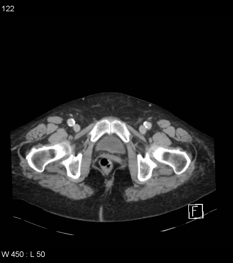
File:Colonic diverticular hemorrhage (Radiopaedia 52561-58475 C 115).jpg
Jump to navigation
Jump to search
Size of this preview: 531 × 599 pixels. Other resolutions: 213 × 240 pixels | 425 × 480 pixels | 766 × 864 pixels.
Original file (766 × 864 pixels, file size: 141 KB, MIME type: image/jpeg)
Summary:
| Description |
|
| Date | Published: 26th Apr 2017 |
| Source | https://radiopaedia.org/cases/colonic-diverticular-haemorrhage |
| Author | Craig Hacking |
| Permission (Permission-reusing-text) |
http://creativecommons.org/licenses/by-nc-sa/3.0/ |
Licensing:
Attribution-NonCommercial-ShareAlike 3.0 Unported (CC BY-NC-SA 3.0)
File history
Click on a date/time to view the file as it appeared at that time.
| Date/Time | Thumbnail | Dimensions | User | Comment | |
|---|---|---|---|---|---|
| current | 16:11, 1 September 2021 | | 766 × 864 (141 KB) | Fæ (talk | contribs) | Radiopaedia project rID:52561 (batch #8718-353 C115) |
You cannot overwrite this file.
File usage
The following page uses this file:
.jpg&oldid=1228014)