File:Congenital diaphragmatic hernia (Radiopaedia 41002).png
Jump to navigation
Jump to search

Size of this preview: 358 × 600 pixels. Other resolutions: 143 × 240 pixels | 286 × 480 pixels | 458 × 768 pixels | 1,104 × 1,849 pixels.
Original file (1,104 × 1,849 pixels, file size: 1,005 KB, MIME type: image/png)
Summary:
- Radiopaedia case ID: 41002
- Image ID: 17336193
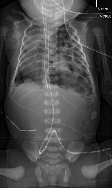
- Study findings: Gastric bubble and colonic loops fill the left hemithorax. Extensive mediastinal shift to the right. Paucity of gas within the abdomen. Umbilical artery catheter at the level of T3. Nasogastric tube tip at the level of the hemidiaphragms. ETT superior to the carina.
- Modality: X-ray
- System: Paediatrics
- Findings: Gastric bubble and colonic loops fill the left hemithorax. Extensive mediastinal shift to the right. Paucity of gas within the abdomen. Umbilical artery catheter at the level of T3. Nasogastric tube tip at the level of the hemidiaphragms. ETT superior to the carina.
- Published: 21st Dec 2015
- Source: https://radiopaedia.org/cases/congenital-diaphragmatic-hernia-10
- Author: Henry Knipe
- Permission: http://creativecommons.org/licenses/by-nc-sa/3.0/
Licensing:
Attribution-NonCommercial-ShareAlike 3.0 Unported (CC BY-NC-SA 3.0)
File history
Click on a date/time to view the file as it appeared at that time.
| Date/Time | Thumbnail | Dimensions | User | Comment | |
|---|---|---|---|---|---|
| current | 19:37, 20 March 2021 | | 1,104 × 1,849 (1,005 KB) | Fæ (talk | contribs) | Radiopaedia project rID:41002 (batch #8652) |
You cannot overwrite this file.
File usage
There are no pages that use this file.
.png&oldid=8858983)