File:Diagram - circumcaval ureter (Radiopaedia 25525).jpg
Jump to navigation
Jump to search
Size of this preview: 600 × 600 pixels. Other resolutions: 240 × 240 pixels | 480 × 480 pixels | 768 × 768 pixels | 1,024 × 1,024 pixels | 1,304 × 1,304 pixels.
Original file (1,304 × 1,304 pixels, file size: 48 KB, MIME type: image/jpeg)
Summary:
- Radiopaedia case ID: 25525
- Image ID: 4931593
- Modality: Diagram
- System: Urogenital
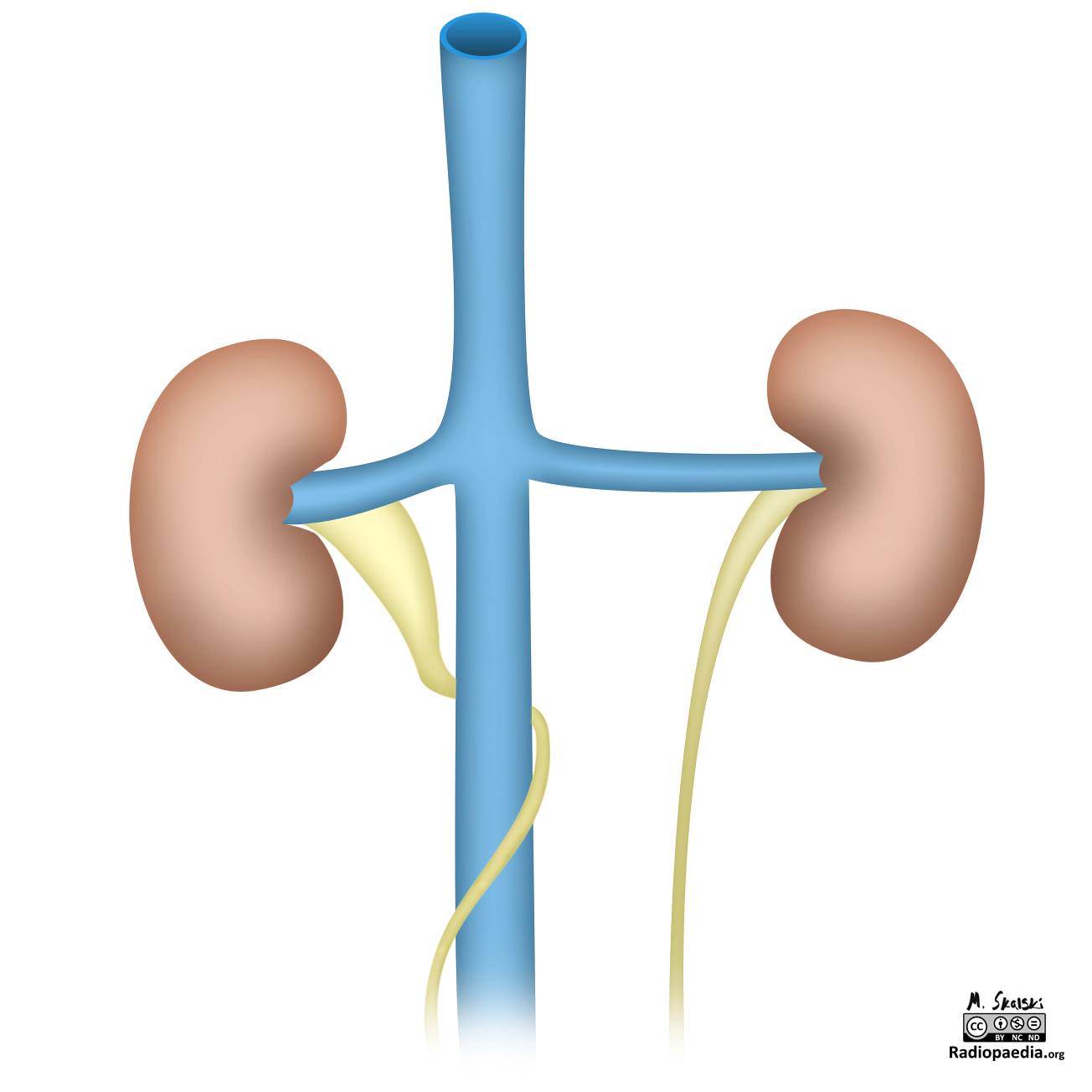
- Findings: The right ureter is depicted passing behind and then around the front of the inferior vena cava, with dilation of the ureter proximal to the retrocaval region.
- Published: 28th Oct 2013
- Source: https://radiopaedia.org/cases/diagram-circumcaval-ureter
- Author: Matt Skalski
- Permission: http://creativecommons.org/licenses/by-nc-sa/3.0/
Licensing:
Attribution-NonCommercial-ShareAlike 3.0 Unported (CC BY-NC-SA 3.0)
| This file is ineligible for copyright and therefore in the public domain, because it is a technical image created as part of a standard medical diagnostic procedure. No creative element rising above the threshold of originality was involved in its production.
|
 |
File history
Click on a date/time to view the file as it appeared at that time.
| Date/Time | Thumbnail | Dimensions | User | Comment | |
|---|---|---|---|---|---|
| current | 12:36, 21 March 2021 | | 1,304 × 1,304 (48 KB) | Fæ (talk | contribs) | Radiopaedia project rID:25525 (batch #10285) |
You cannot overwrite this file.
File usage
There are no pages that use this file.
.jpg&oldid=9754143)