File:Diagram of the ductal anatomy of the breast.png
Jump to navigation
Jump to search
No higher resolution available.
Diagram_of_the_ductal_anatomy_of_the_breast.png (512 × 596 pixels, file size: 518 KB, MIME type: image/png)
Summary
| Description |
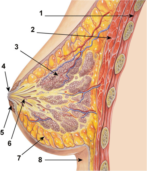
English: Diagram of the ductal anatomy of the breast. (1) Chest wall, (2) Pectoral muscles, (3) Lobules, (4) Nipple surface, (5) Areola, (6) Lactiferous duct, (7) Fatty tissue, (8) Skin (original author: Patrick J. Lynch; reworked by Morgoth666 to add numbered legend. (Patrick J. Lynch, medical illustrator) [CC-BY-3.0 (http://creativecommons.org/licenses/by/3.0)], via Wikimedia Commons). |
| Date | |
| Source | In vivo assessment of number of milk duct orifices in lactating women and association with parameters in the mother and the infant. BMC Pregnancy Childbirth 14, 124 (2014). https://doi.org/10.1186/1471-2393-14-124. Via Openi |
| Author | Jütte, J., Hohoff, A., Sauerland, C. et al. |
Licensing
{{subst:Custom license marker added by UW}} CC BY 2.0 DEED
This file was uploaded with UploadWizard.
File history
Click on a date/time to view the file as it appeared at that time.
| Date/Time | Thumbnail | Dimensions | User | Comment | |
|---|---|---|---|---|---|
| current | 06:28, 26 January 2024 | | 512 × 596 (518 KB) | Whispyhistory (talk | contribs) | Uploaded a work by Jütte, J., Hohoff, A., Sauerland, C. et al. from [https://bmcpregnancychildbirth.biomedcentral.com/articles/10.1186/1471-2393-14-124 In vivo assessment of number of milk duct orifices in lactating women and association with parameters in the mother and the infant]. BMC Pregnancy Childbirth 14, 124 (2014). https://doi.org/10.1186/1471-2393-14-124. [https://openi.nlm.nih.gov/detailedresult?img=PMC3992155_1471-2393-14-124-2&query=&req=4 Via Openi] with UploadWizard |
You cannot overwrite this file.
File usage
There are no pages that use this file.