File:Duplicated middle cerebral artery (Radiopaedia 6095).jpg
Jump to navigation
Jump to search

Size of this preview: 600 × 600 pixels. Other resolutions: 240 × 240 pixels | 480 × 480 pixels | 768 × 768 pixels | 1,024 × 1,024 pixels | 1,280 × 1,280 pixels.
Original file (1,280 × 1,280 pixels, file size: 219 KB, MIME type: image/jpeg)
Summary:
- Radiopaedia case ID: 6095
- Image ID: 24360
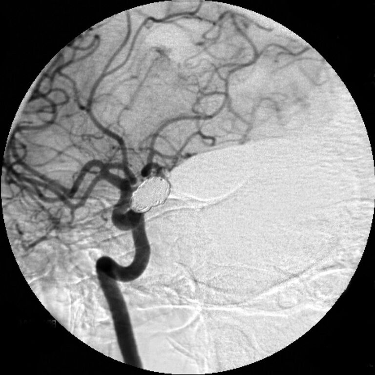
- Study findings: This patient's middle cerebral artery is duplicated, or if you prefer the M1 segment is very short (non-existent) and the bifurcation occurs at the terminal internal carotid artery. This condition is named as middle cerebral artery duplication. This must be differentiated from another variant in which the anomalous vessel originates from the anterior cerebral artery, named as the accessory middle cerebral artery.
- Modality: DSA (angiography)
- System: Vascular
- Findings: This patient's middle cerebral artery is duplicated, or if you prefer the M1 segment is very short (non-existent) and the bifurcation occurs at the terminal internal carotid artery. This condition is named as middle cerebral artery duplication. This must be differentiated from another variant in which the anomalous vessel originates from the anterior cerebral artery, named as the accessory middle cerebral artery.
- Published: 24th Apr 2009
- Source: https://radiopaedia.org/cases/duplicated-middle-cerebral-artery-2
- Author: Frank Gaillard
- Permission: http://creativecommons.org/licenses/by-nc-sa/3.0/
Licensing:
Attribution-NonCommercial-ShareAlike 3.0 Unported (CC BY-NC-SA 3.0)
File history
Click on a date/time to view the file as it appeared at that time.
| Date/Time | Thumbnail | Dimensions | User | Comment | |
|---|---|---|---|---|---|
| current | 17:44, 21 March 2021 | | 1,280 × 1,280 (219 KB) | Fæ (talk | contribs) | Radiopaedia project rID:6095 (batch #11301) |
You cannot overwrite this file.
File usage
There are no pages that use this file.
.jpg&oldid=8858532)