File:EAEC (1).jpg
Jump to navigation
Jump to search
No higher resolution available.
EAEC_(1).jpg (256 × 256 pixels, file size: 8 KB, MIME type: image/jpeg)
Summary
| Description |
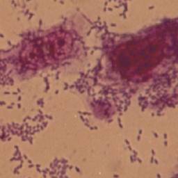
English: Aggregative adherence pattern of EAEC. HEp-2 cells (106) were grown to 50%–70% confluency as monolayers in a 6-well flat-bottom tissue culture plate. After overnight growth cells were washed and 2 mL of fresh DMEM media (pH 7.4) was added, and EAEC grown overnight at 37°C (215 rpm) was inoculated (25 μL) into the plate and incubated at 37°C overnight in 5% CO2 atmosphere. Following incubation, the cells were washed, fixed, and stained with 2.5% Giemsa for 15 minutes. The adherence patterns were examined under 40-X magnification and photographed at 100-X magnification with digital camera in a light microscope. Data are a representative experiment from three independently performed experiments with similar results. |
| Date | |
| Source | https://www.ncbi.nlm.nih.gov/pmc/articles/PMC2837894/ |
| Author | P. Kaur,A. Chakraborti, A. Asea |
Licensing
English: This file is licensed CC BY-NC 4.0
This file was uploaded with UploadWizard.
File history
Click on a date/time to view the file as it appeared at that time.
| Date/Time | Thumbnail | Dimensions | User | Comment | |
|---|---|---|---|---|---|
| current | 19:36, 21 August 2022 | | 256 × 256 (8 KB) | Ozzie10aaaa (talk | contribs) | Uploaded a work by P. Kaur,A. Chakraborti, A. Asea from https://www.ncbi.nlm.nih.gov/pmc/articles/PMC2837894/ with UploadWizard |
You cannot overwrite this file.
File usage
There are no pages that use this file.
.jpg&oldid=1253938)