File:Endosalpingiosis1.png
Jump to navigation
Jump to search
No higher resolution available.
Endosalpingiosis1.png (512 × 340 pixels, file size: 341 KB, MIME type: image/png)
Summary
| Description |
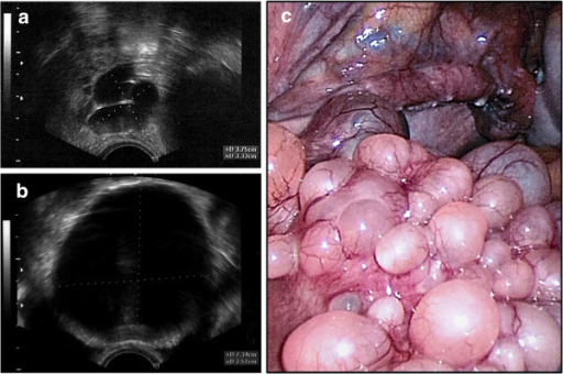
English: Transvaginal ultrasound showed cystic formations in the areas of the right fallopian tube (a) and fundus uteri (b). The maximum diameter of the cysts was 7.5 cm. Laparoscopic presentation of the cystic mass (c); uterus (front) with right tube and right abdominal wall (background). |
| Date | |
| Source | Cystic endosalpingiosis presenting as chronic back pain, a case report. Diagn Pathol. 2013 Dec 3;8:196. doi: 10.1186/1746-1596-8-196. PMID: 24299296; PMCID: PMC3924907. ViaOpeni |
| Author | Scheel AH, Frasunek J, Meyer W, Ströbel P. |
Licensing
{{subst:Custom license marker added by UW}} Attribution 2.0 Generic (CC BY 2.0)
This file was uploaded with UploadWizard.
File history
Click on a date/time to view the file as it appeared at that time.
| Date/Time | Thumbnail | Dimensions | User | Comment | |
|---|---|---|---|---|---|
| current | 10:23, 29 July 2022 | | 512 × 340 (341 KB) | Whispyhistory (talk | contribs) | Uploaded a work by Scheel AH, Frasunek J, Meyer W, Ströbel P. from [https://www.ncbi.nlm.nih.gov/pmc/articles/PMC3924907/ Cystic endosalpingiosis presenting as chronic back pain, a case report]. Diagn Pathol. 2013 Dec 3;8:196. doi: 10.1186/1746-1596-8-196. PMID: 24299296; PMCID: PMC3924907. [https://openi.nlm.nih.gov/detailedresult?img=PMC3924907_1746-1596-8-196-1&query=Endosalpingiosis&it=xg&req=4&npos=36 ViaOpeni] with UploadWizard |
You cannot overwrite this file.
File usage
There are no pages that use this file.