File:Ependymitis granularis (Radiopaedia 10904).jpg
Jump to navigation
Jump to search
Size of this preview: 522 × 599 pixels. Other resolutions: 209 × 240 pixels | 418 × 480 pixels | 766 × 879 pixels.
Original file (766 × 879 pixels, file size: 68 KB, MIME type: image/jpeg)
Summary:
- Radiopaedia case ID: 10904
- Image ID: 550484
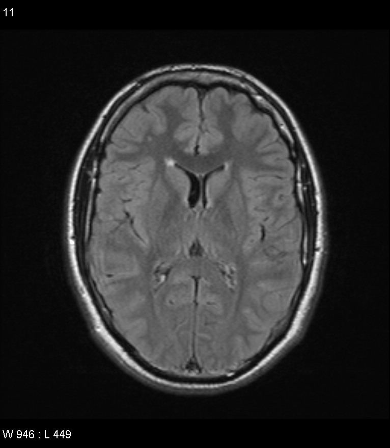
- Modality: MRI
- System: Central Nervous System
- Findings: Single FLAIR axial image through the frontal horns of the lateral ventricles demonstrates small triangular regions of high signal abutting the anterolateral aspect of the ventricles. This is a normal finding which is called ependymitis granularis.
- Published: 30th Sep 2010
- Source: https://radiopaedia.org/cases/ependymitis-granularis-1
- Author: Paul Smith
- Permission: http://creativecommons.org/licenses/by-nc-sa/3.0/
Licensing:
Attribution-NonCommercial-ShareAlike 3.0 Unported (CC BY-NC-SA 3.0)
File history
Click on a date/time to view the file as it appeared at that time.
| Date/Time | Thumbnail | Dimensions | User | Comment | |
|---|---|---|---|---|---|
| current | 20:10, 21 March 2021 | | 766 × 879 (68 KB) | Fæ (talk | contribs) | Radiopaedia project rID:10904 (batch #12156) |
You cannot overwrite this file.
File usage
There are no pages that use this file.
.jpg&oldid=8858431)