File:Ependymoma (histology) (Radiopaedia 51845).JPG
Jump to navigation
Jump to search

Size of this preview: 600 × 600 pixels. Other resolutions: 240 × 240 pixels | 480 × 480 pixels | 768 × 768 pixels | 1,163 × 1,163 pixels.
Original file (1,163 × 1,163 pixels, file size: 1.3 MB, MIME type: image/jpeg)
Summary:
- Radiopaedia case ID: 51845
- Image ID: 29100640
- Modality: Pathology
- System: Central Nervous System
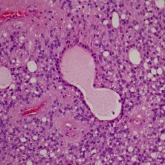
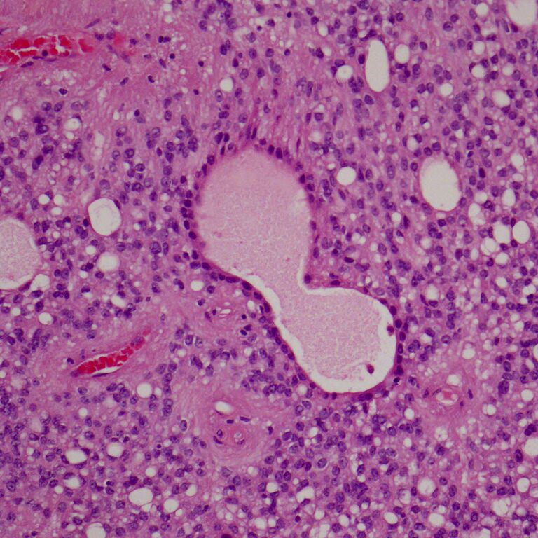
- Findings: H&E section through an ependymoma demonstrating a "figure 8" ependymal rosette (or ependymal canals); cuboidal tumor cells surrounding an empty lumen. They are far less common than perivascular pseudorosettes (a small perivascular pseudorosette is seen just below and to the left of the true rosette.
- Published: 10th Mar 2017
- Source: https://radiopaedia.org/cases/ependymoma-histology-1
- Author: Frank Gaillard
- Permission: http://creativecommons.org/licenses/by-nc-sa/3.0/
Licensing:
Attribution-NonCommercial-ShareAlike 3.0 Unported (CC BY-NC-SA 3.0)
File history
Click on a date/time to view the file as it appeared at that time.
| Date/Time | Thumbnail | Dimensions | User | Comment | |
|---|---|---|---|---|---|
| current | 20:16, 21 March 2021 | | 1,163 × 1,163 (1.3 MB) | Fæ (talk | contribs) | Radiopaedia project rID:51845 (batch #12189) |
You cannot overwrite this file.
File usage
There are no pages that use this file.
_(Radiopaedia_51845).JPG&oldid=8858428)