File:Fibroadenoma calcifications (Radiopaedia 18921).jpg
Jump to navigation
Jump to search
Size of this preview: 383 × 600 pixels. Other resolutions: 153 × 240 pixels | 306 × 480 pixels | 490 × 768 pixels | 654 × 1,024 pixels | 1,512 × 2,367 pixels.
Original file (1,512 × 2,367 pixels, file size: 379 KB, MIME type: image/jpeg)
Summary:
- Radiopaedia case ID: 18921
- Image ID: 2189662
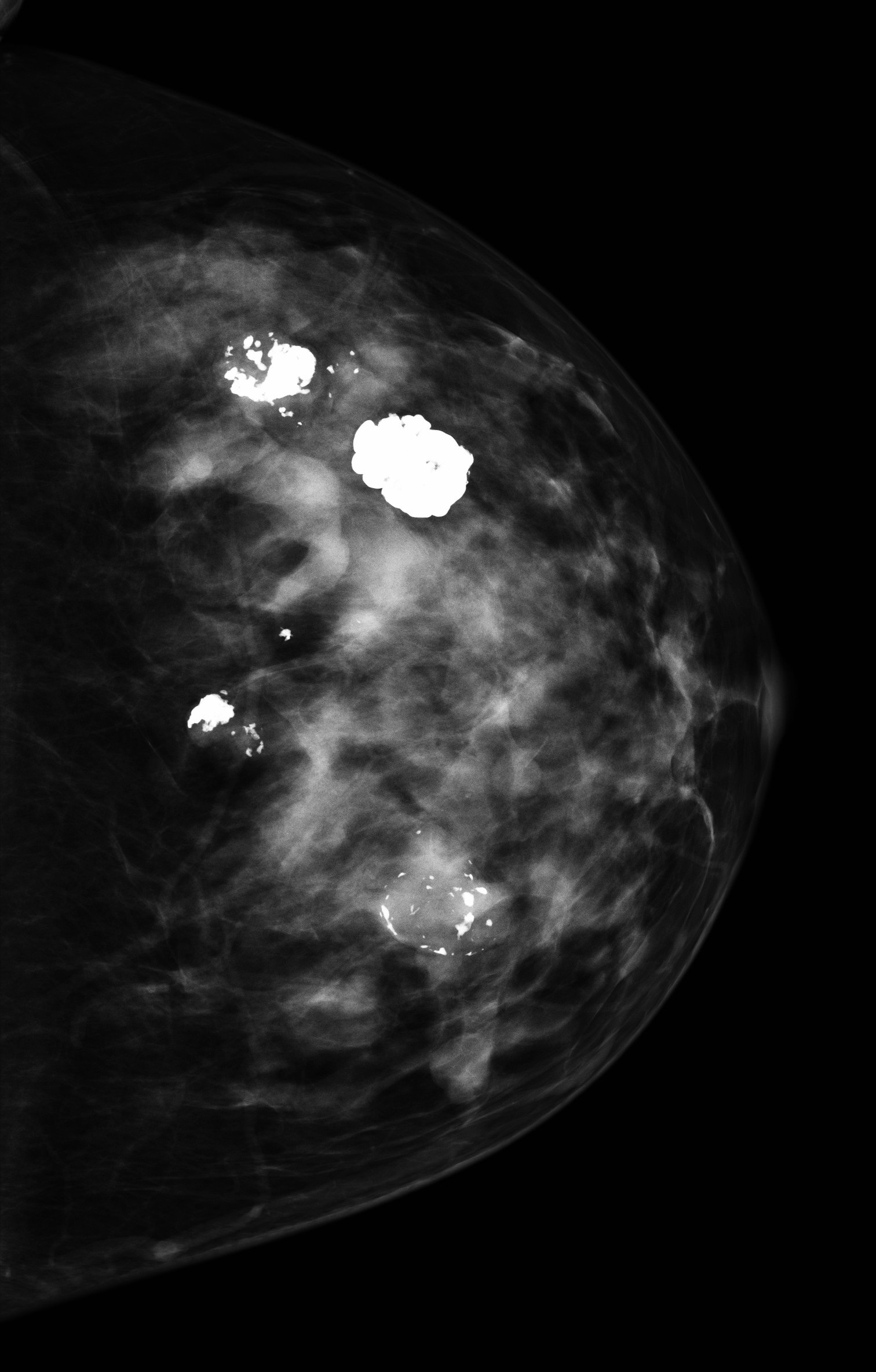
- Study findings: The cc image shows the spectrum of fibroadenoma calcifications in one breast. The density laterally (i.e at the top of the image) with no calcium is a fibroadenoma. The calcification then starts peripherally in the medial (bottom) lesion and progresses in the central lesion to ultimately complete calcify the lesion lateral anterior.
- Modality: Mammography
- System: Breast
- Findings: The cc image shows the spectrum of fibroadenoma calcifications in one breast. The density laterally (i. e at the top of the image) with no calcium is a fibroadenoma. The calcification then starts peripherally in the medial (bottom) lesion and progresses in the central lesion to ultimately complete calcify the lesion lateral anterior.
- Published: 27th Jul 2012
- Source: https://radiopaedia.org/cases/fibroadenoma-calcifications
- Author: Garth Kruger
- Permission: http://creativecommons.org/licenses/by-nc-sa/3.0/
Licensing:
Attribution-NonCommercial-ShareAlike 3.0 Unported (CC BY-NC-SA 3.0)
File history
Click on a date/time to view the file as it appeared at that time.
| Date/Time | Thumbnail | Dimensions | User | Comment | |
|---|---|---|---|---|---|
| current | 01:07, 22 March 2021 | | 1,512 × 2,367 (379 KB) | Fæ (talk | contribs) | Radiopaedia project rID:18921 (batch #13302) |
You cannot overwrite this file.
File usage
There are no pages that use this file.
.jpg&oldid=8854399)