File:Fibrolipomatous hamartoma of median nerve (Radiopaedia 7825).jpg
Jump to navigation
Jump to search
No higher resolution available.
Fibrolipomatous_hamartoma_of_median_nerve_(Radiopaedia_7825).jpg (460 × 430 pixels, file size: 18 KB, MIME type: image/jpeg)
Summary:
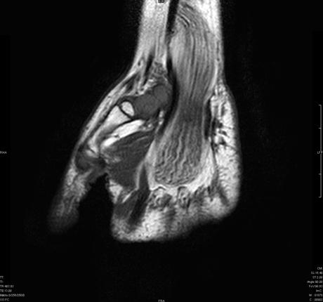
- Radiopaedia case ID: 7825
- Image ID: 4483
- Modality: MRI
- System: Musculoskeletal
- Findings: MR imaging demonstrates fusiform median nerve enlargement which is caused by thickening of nerve bundles and fatty and fibrous proliferation. The serpiginous low-intensity structures represent thickened nerve fascicles, surrounded by evenly distributed fat of high signal intensity on this T1-weighted image. A spaghetti-like appearance is seen in the coronal plane. The median nerve is the most commonly involved nerve The MR imaging characteristics of fibrolipomatous hamartoma are pathognomonic, obviating the need for biopsy for diagnosis.
- Published: 15th Dec 2009
- Source: https://radiopaedia.org/cases/fibrolipomatous-hamartoma-of-median-nerve
- Author: Frank Gaillard
- Permission: http://creativecommons.org/licenses/by-nc-sa/3.0/
Licensing:
Attribution-NonCommercial-ShareAlike 3.0 Unported (CC BY-NC-SA 3.0)
File history
Click on a date/time to view the file as it appeared at that time.
| Date/Time | Thumbnail | Dimensions | User | Comment | |
|---|---|---|---|---|---|
| current | 01:11, 22 March 2021 | | 460 × 430 (18 KB) | Fæ (talk | contribs) | Radiopaedia project rID:7825 (batch #13327) |
You cannot overwrite this file.
File usage
There are no pages that use this file.
.jpg&oldid=8858159)