File:Fibular hemimelia (Radiopaedia 48168).png
Jump to navigation
Jump to search
Fibular_hemimelia_(Radiopaedia_48168).png (427 × 326 pixels, file size: 119 KB, MIME type: image/png)
Summary:
- Radiopaedia case ID: 48168
- Image ID: 25245523
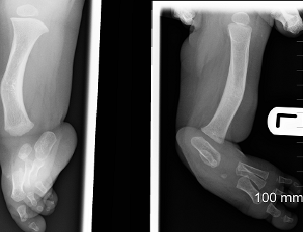
- Study findings: The left tibia is foreshortened, thickened and bowed anteromedially. The left fibula is absent. There is evidence of talipes equinovalgus deformity. The lateral rays and phalanges (4th and 5th digits) are also absent. Findings are consistent with type II Fibular hemimelia.
- Modality: X-ray
- System: Paediatrics
- Findings: The left tibia is foreshortened, thickened and bowed anteromedially. The left fibula is absent. There is evidence of talipes equinovalgus deformity. The lateral rays and phalanges (4th and 5th digits) are also absent. Findings are consistent with type II Fibular hemimelia.
- Published: 23rd Sep 2016
- Source: https://radiopaedia.org/cases/fibular-hemimelia-2
- Author: Yusra Sheikh
- Permission: http://creativecommons.org/licenses/by-nc-sa/3.0/
Licensing:
Attribution-NonCommercial-ShareAlike 3.0 Unported (CC BY-NC-SA 3.0)
File history
Click on a date/time to view the file as it appeared at that time.
| Date/Time | Thumbnail | Dimensions | User | Comment | |
|---|---|---|---|---|---|
| current | 01:30, 22 March 2021 |  | 427 × 326 (119 KB) | Fæ (talk | contribs) | Radiopaedia project rID:48168 (batch #13444) |
You cannot overwrite this file.
File usage
There are no pages that use this file.
