File:Fnhum-11-00103-g001.jpg
Jump to navigation
Jump to search

Size of this preview: 485 × 599 pixels. Other resolutions: 194 × 240 pixels | 388 × 480 pixels | 1,000 × 1,236 pixels.
Original file (1,000 × 1,236 pixels, file size: 232 KB, MIME type: image/jpeg)
Summary
| Description |
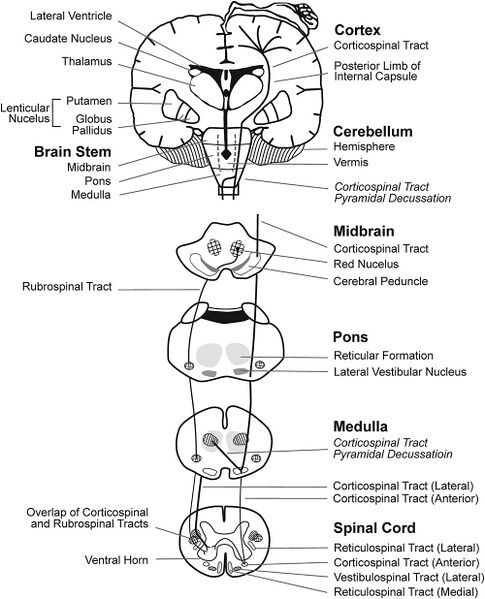
English: Brain regions and WM motor tracts affected in spastic, dyskinetic, and ataxic CP. A portion of the motor homunculus is superimposed on the cortex. Representations of the lateral ventricles, subcortical nuclei, cerebellum, brain stem, and rubrospinal and CSTs are outlined. The vermis of the cerebellum (posterior to the brainstem) is represented by dotted lines. Regions where the reticulospinal and vestibulospinal tracts descend through each layer of the brainstem and the spinal cord are shaded. |
| Date | |
| Source | https://www.ncbi.nlm.nih.gov/pmc/articles/PMC5355477/ |
| Author | Joanne Zhou,1,2 Erin E. Butler,3,4 and Jessica Rose1 |
Licensing
English: This file is licensed CC BY-NC 4.0
This file was uploaded with UploadWizard.
File history
Click on a date/time to view the file as it appeared at that time.
| Date/Time | Thumbnail | Dimensions | User | Comment | |
|---|---|---|---|---|---|
| current | 22:05, 12 February 2023 | | 1,000 × 1,236 (232 KB) | Ozzie10aaaa (talk | contribs) | Uploaded a work by Joanne Zhou,1,2 Erin E. Butler,3,4 and Jessica Rose1 from https://www.ncbi.nlm.nih.gov/pmc/articles/PMC5355477/ with UploadWizard |
You cannot overwrite this file.
File usage
There are no pages that use this file.