File:Focal fatty change (Radiopaedia 7935).jpg
Jump to navigation
Jump to search
No higher resolution available.
Focal_fatty_change_(Radiopaedia_7935).jpg (456 × 600 pixels, file size: 84 KB, MIME type: image/jpeg)
Summary:
- Radiopaedia case ID: 7935
- Image ID: 728
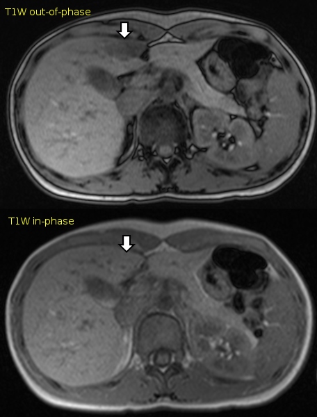
- Study findings: These in- and out-of-phase T1-weighted gradient-echo sequences show focal fat infiltration to the right of the fissure for the ligamentum teres. There is signal loss on out-of-phase images. Signal loss occurs when water and fat molecules occupy the same voxel, and the signals they return negatively interfere. This is due to the different precession speeds of fat and water in the same magnetic field. At 1.5 T, the water and fat signal are in phase when TE is an even multiple, and out of phase when TE is an odd multiple of 2.3 ms.
- Modality: MRI
- System: Gastrointestinal
- Findings: These in- and out-of-phase T1-weighted gradient-echo sequences show focal fat infiltration to the right of the fissure for the ligamentum teres. There is signal loss on out-of-phase images. Signal loss occurs when water and fat molecules occupy the same voxel, and the signals they return negatively interfere. This is due to the different precession speeds of fat and water in the same magnetic field. At 1. 5 T, the water and fat signal are in phase when TE is an even multiple, and out of phase when TE is an odd multiple of 2. 3 ms.
- Published: 24th Dec 2009
- Source: https://radiopaedia.org/cases/focal-fatty-change
- Author: Frank Gaillard
- Permission: http://creativecommons.org/licenses/by-nc-sa/3.0/
Licensing:
Attribution-NonCommercial-ShareAlike 3.0 Unported (CC BY-NC-SA 3.0)
File history
Click on a date/time to view the file as it appeared at that time.
| Date/Time | Thumbnail | Dimensions | User | Comment | |
|---|---|---|---|---|---|
| current | 09:27, 22 March 2021 | | 456 × 600 (84 KB) | Fæ (talk | contribs) | Radiopaedia project rID:7935 (batch #13653) |
You cannot overwrite this file.
File usage
There are no pages that use this file.
.jpg&oldid=8858094)