File:Gastrointestinal stromal tumor (gross pathology) (Radiopaedia 36400).jpg
Jump to navigation
Jump to search

Size of this preview: 600 × 600 pixels. Other resolutions: 240 × 240 pixels | 480 × 480 pixels | 768 × 768 pixels | 1,024 × 1,024 pixels | 1,600 × 1,600 pixels.
Original file (1,600 × 1,600 pixels, file size: 211 KB, MIME type: image/jpeg)
Summary:
- Radiopaedia case ID: 36400
- Image ID: 27931
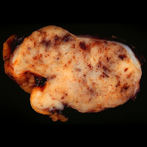
- Study findings: GIST of the stomach. In this photo, the gastric wall with the ulcer is on the left side of the image. Note how well the mucosa is demarcated from the tumor surface. Tags of dark liver tissue, barely visible, adhere to the tumor on the right side of the image.
- Modality: Pathology
- System: Gastrointestinal
- Findings: GIST of the stomach. In this photo, the gastric wall with the ulcer is on the left side of the image. Note how well the mucosa is demarcated from the tumor surface. Tags of dark liver tissue, barely visible, adhere to the tumor on the right side of the image.
- Published: 18th May 2015
- Source: https://radiopaedia.org/cases/gastrointestinal-stromal-tumour-gross-pathology-2
- Author: Ed Uthman
- Permission: http://creativecommons.org/licenses/by-nc-sa/3.0/
Licensing:
Attribution-NonCommercial-ShareAlike 3.0 Unported (CC BY-NC-SA 3.0)
| This file is ineligible for copyright and therefore in the public domain, because it is a technical image created as part of a standard medical diagnostic procedure. No creative element rising above the threshold of originality was involved in its production.
|
 |
File history
Click on a date/time to view the file as it appeared at that time.
| Date/Time | Thumbnail | Dimensions | User | Comment | |
|---|---|---|---|---|---|
| current | 12:37, 22 March 2021 | | 1,600 × 1,600 (211 KB) | Fæ (talk | contribs) | Radiopaedia project rID:36400 (batch #14786) |
You cannot overwrite this file.
File usage
There are no pages that use this file.
_(Radiopaedia_36400).jpg&oldid=9755300)