File:Hydatid cyst of liver (gross pathology) (Radiopaedia 29673).jpg
Jump to navigation
Jump to search

Size of this preview: 600 × 600 pixels. Other resolutions: 240 × 240 pixels | 480 × 480 pixels.
Original file (800 × 800 pixels, file size: 213 KB, MIME type: image/jpeg)
Summary:
- Radiopaedia case ID: 29673
- Image ID: 7381049
- Modality: Pathology
- System: Hepatobiliary
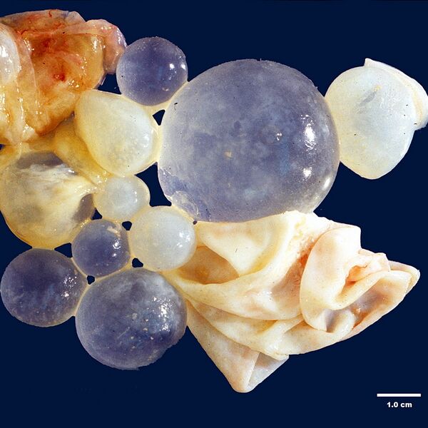
- Findings: Abdominal CT scan (not shown) showed a single large cyst in the right lobe of liver measuring 12 cm. It appeared to contain several smaller cysts within it. The findings were considered to be diagnostic of hydatid cyst. The patient also had mild peripheral eosinophilia. The image shows parts of the collapsed wall of the larger cyst (lower right and upper left) and several intact daughter cysts ranging in size from 1 cm to 5 cm.
- Published: 12th Jun 2014
- Source: https://radiopaedia.org/cases/hydatid-cyst-of-liver-gross-pathology-1
- Author: Dharam Ramnani
- Permission: http://creativecommons.org/licenses/by-nc-sa/3.0/
Licensing:
Attribution-NonCommercial-ShareAlike 3.0 Unported (CC BY-NC-SA 3.0)
| This file is ineligible for copyright and therefore in the public domain, because it is a technical image created as part of a standard medical diagnostic procedure. No creative element rising above the threshold of originality was involved in its production.
|
 |
File history
Click on a date/time to view the file as it appeared at that time.
| Date/Time | Thumbnail | Dimensions | User | Comment | |
|---|---|---|---|---|---|
| current | 20:44, 22 March 2021 | | 800 × 800 (213 KB) | Fæ (talk | contribs) | Radiopaedia project rID:29673 (batch #16924) |
You cannot overwrite this file.
File usage
There are no pages that use this file.
_(Radiopaedia_29673).jpg&oldid=9756262)