File:Images large 10.1177 20420188211058323-fig1 (1).jpg
Jump to navigation
Jump to search

Size of this preview: 800 × 449 pixels. Other resolutions: 320 × 179 pixels | 640 × 359 pixels | 1,024 × 574 pixels | 1,789 × 1,003 pixels.
Original file (1,789 × 1,003 pixels, file size: 577 KB, MIME type: image/jpeg)
Summary
| Description |
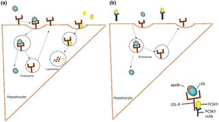
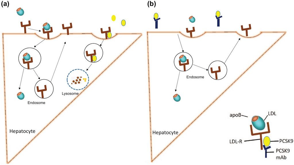
English: Figure 1. The mechanism of action of PCSK9 inhibitors. (a) Shows that LDL-C attaches to LDL-R to incorporate it into the hepatocyte. This can either be recycled when not attached to PCSK9. When the secreted PCSK9 attaches to the LDL-R, it eventually leads to lysosomal degradation of PCSK9. (b) Illustrates that in the presence of PCSK9 monoclonal antibody, the PCSK9 is inactivated, preventing the LDL-R from being degraded. This allows for the LDL-R to be recycled, prompting LDL-C uptake, and reducing LDL-C levels in the serum. |
| Date | |
| Source | https://journals.sagepub.com/doi/10.1177/20420188211058323 |
| Author | Bhuvana Sunil, Christy Foster, Ambika P. Ashraf |
Licensing
English: This file is licensed CC BY-NC 4.0
This file was uploaded with UploadWizard.
File history
Click on a date/time to view the file as it appeared at that time.
| Date/Time | Thumbnail | Dimensions | User | Comment | |
|---|---|---|---|---|---|
| current | 17:30, 5 May 2024 | | 1,789 × 1,003 (577 KB) | Ozzie10aaaa (talk | contribs) | Uploaded a work by Bhuvana Sunil, Christy Foster, Ambika P. Ashraf from https://journals.sagepub.com/doi/10.1177/20420188211058323 with UploadWizard |
You cannot overwrite this file.
File usage
There are no pages that use this file.
.jpg&oldid=9797637)