File:Intracranial hypotension (Radiopaedia 36019).jpg
Jump to navigation
Jump to search
Size of this preview: 566 × 600 pixels. Other resolutions: 227 × 240 pixels | 453 × 480 pixels | 995 × 1,054 pixels.
Original file (995 × 1,054 pixels, file size: 245 KB, MIME type: image/jpeg)
Summary:
- Radiopaedia case ID: 36019
- Image ID: 1030
- Modality: MRI
- System: Central Nervous System
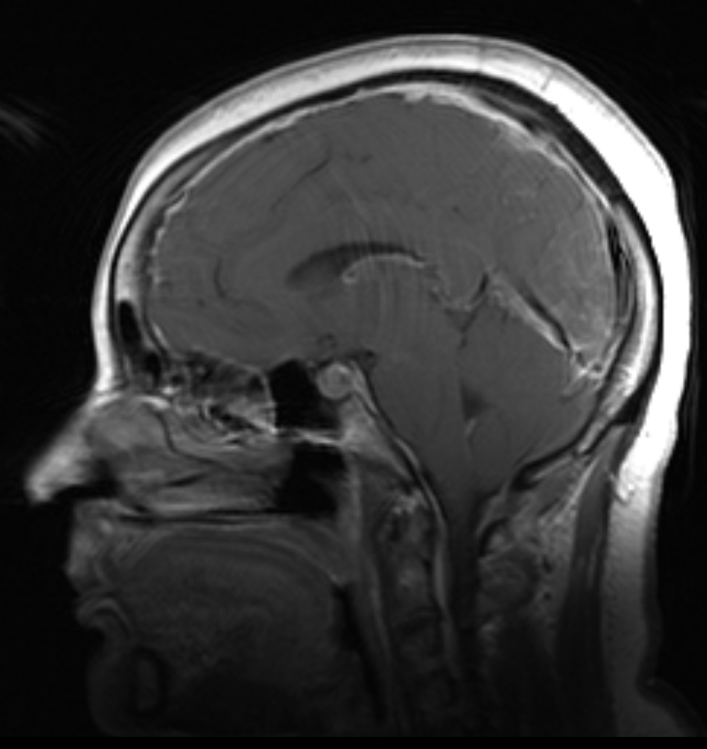
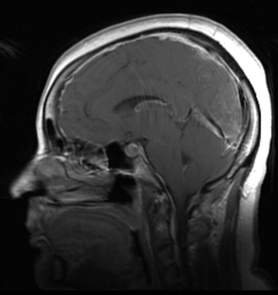
- Findings: Midsagittal contrast enhanced MRI of the brain in a 60-year-old woman with postural headache shows smooth pachymeningeal enhancement, effacement of cortical sulci (more than would be expected for a 60-year-old person), sagging cerebellar tonsils, and a plump, full pituitary gland. These findings are consistent with intracranial hypotension.
- Published: 12th May 2015
- Source: https://radiopaedia.org/cases/intracranial-hypotension-2
- Author: Behrang Amini
- Permission: http://creativecommons.org/licenses/by-nc-sa/3.0/
Licensing:
Attribution-NonCommercial-ShareAlike 3.0 Unported (CC BY-NC-SA 3.0)
| This file is ineligible for copyright and therefore in the public domain, because it is a technical image created as part of a standard medical diagnostic procedure. No creative element rising above the threshold of originality was involved in its production.
|
 |
File history
Click on a date/time to view the file as it appeared at that time.
| Date/Time | Thumbnail | Dimensions | User | Comment | |
|---|---|---|---|---|---|
| current | 13:56, 23 March 2021 | | 995 × 1,054 (245 KB) | Fæ (talk | contribs) | Radiopaedia project rID:36019 (batch #18358) |
You cannot overwrite this file.
File usage
There are no pages that use this file.
.jpg&oldid=9756162)