File:Jem2040595f01 (1) (1).png
Jump to navigation
Jump to search
No higher resolution available.
Jem2040595f01_(1)_(1).png (128 × 144 pixels, file size: 36 KB, MIME type: image/png)
Summary
| Description |
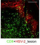
English: fig1: Spatial and functional CD8+ CTL activity during an acute genital herpetic lesion. (a) Hematoxylin and eosin staining of normal arm (left) and lesional skin (middle). Immunofluorescence staining (right) for CD8+ cells (green) and HSV-2–infected cells (red). (b) Confocal image of CD8+ and HSV-2–infected cells in an acute lesion. Montages of a CD8+ cell (green) in proximity to HSV-2–infected cells (blue) were displayed at 1-μm intervals (left). (middle and right) The 0° (front) and 180° (back) views of a three-dimensional rendering of the interactions. (c) HSV-2–specific CD8 cytolytic activity as measured by chromium release in lymphocytes cultured from a lesion biopsy 5 d after onset, using either bulk or CD8-enriched cultured cells as effectors, and either the autologous or HLA-mismatched B lymphocyte blasts uninfected or HSV-2 infected as targets. Effector/target cell ratio = 20:1. Bars: (a) 50 μm; (b) 10 μm. |
| Date | |
| Source | https://openi.nlm.nih.gov/detailedresult?img=PMC2137910_jem2040595f01&query=Human%20alphaherpesvirus%202&it=xg&req=4&npos=11 |
| Author | Zhu J, Koelle DM, Cao J, Vazquez J, Huang ML, Hladik F, Wald A, Corey L |
Licensing
Creative Commons Attribution-NonCommercial-ShareAlike 4.0 International
This file was uploaded with UploadWizard.
File history
Click on a date/time to view the file as it appeared at that time.
| Date/Time | Thumbnail | Dimensions | User | Comment | |
|---|---|---|---|---|---|
| current | 18:54, 10 May 2023 | | 128 × 144 (36 KB) | Ozzie10aaaa (talk | contribs) | Uploaded a work by Zhu J, Koelle DM, Cao J, Vazquez J, Huang ML, Hladik F, Wald A, Corey L from https://openi.nlm.nih.gov/detailedresult?img=PMC2137910_jem2040595f01&query=Human%20alphaherpesvirus%202&it=xg&req=4&npos=11 with UploadWizard |
You cannot overwrite this file.
File usage
There are no pages that use this file.
_(1).png&oldid=1252678)