File:Leukoencephalopathy with calcifications and cysts (histology) (Radiopaedia 60264).png
Jump to navigation
Jump to search

Size of this preview: 800 × 600 pixels. Other resolutions: 320 × 240 pixels | 640 × 480 pixels | 1,024 × 768 pixels | 1,280 × 960 pixels | 1,600 × 1,200 pixels.
Original file (1,600 × 1,200 pixels, file size: 3.08 MB, MIME type: image/png)
Summary:
- Radiopaedia case ID: 60264
- Image ID: 38822265
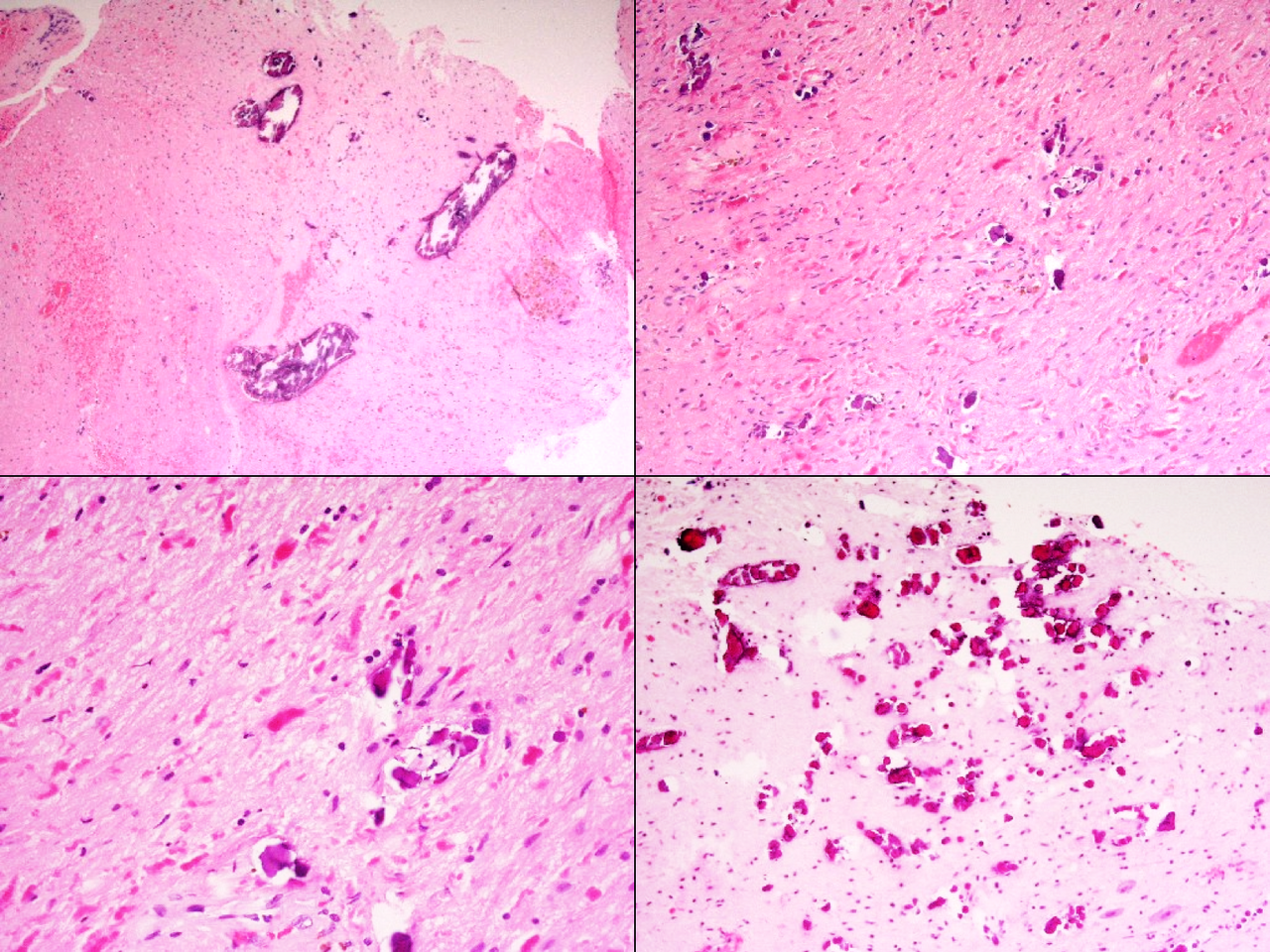
- Study findings: Histology of a patient with leukoencephalopathy with calcifications and cysts demonstrating a combination of calcification and gliosis with exuberant Rosenthal fiber formation. The calcification can be linear alongside blood vessels (upper left panel) and therefore can mimic Fahr’s disease. Histology courtesy of Professor Michael Gonzales, Royal Melbourne Hospital, Australia.
- Modality: Pathology
- System: Central Nervous System
- Findings: Histology of a patient with leukoencephalopathy with calcifications and cysts demonstrating a combination of calcification and gliosis with exuberant Rosenthal fiber formation. The calcification can be linear alongside blood vessels (upper left panel) and therefore can mimic Fahr’s disease. Histology courtesy of Professor Michael Gonzales, Royal Melbourne Hospital, Australia.
- Published: 14th May 2018
- Source: https://radiopaedia.org/cases/leukoencephalopathy-with-calcifications-and-cysts-histology
- Author: Frank Gaillard
- Permission: http://creativecommons.org/licenses/by-nc-sa/3.0/
Licensing:
Attribution-NonCommercial-ShareAlike 3.0 Unported (CC BY-NC-SA 3.0)
File history
Click on a date/time to view the file as it appeared at that time.
| Date/Time | Thumbnail | Dimensions | User | Comment | |
|---|---|---|---|---|---|
| current | 05:01, 24 March 2021 | | 1,600 × 1,200 (3.08 MB) | Fæ (talk | contribs) | Radiopaedia project rID:60264 (batch #20316) |
You cannot overwrite this file.
File usage
There are no pages that use this file.
_(Radiopaedia_60264).png&oldid=8856707)