File:Medi-96-e7365-g001.jpg
Jump to navigation
Jump to search

Size of this preview: 732 × 599 pixels. Other resolutions: 293 × 240 pixels | 586 × 480 pixels.
Original file (800 × 655 pixels, file size: 130 KB, MIME type: image/jpeg)
Summary
| Description |
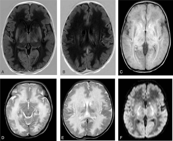
English: Brain magnetic resonance imaging (MRI) on day 18 of life. MRI revealed extensive abnormalities in the deep white matter of the bilateral cerebral hemisphere, subcortical white matter, caudate nuclei, the dorsal thalamus, and the cerebellar hemisphere, which suggested hereditary metabolic leukoencephalopathy. (A and B) Low signal intensity on T1 weighted image (T1WI); (C) high signal intensity on fluid attenuated inversion recovery (FLAIR); (D and E) high signal intensity on T2 weighted image (T2WI); (F) high signal intensity on diffusion weighted image (DWI). |
| Date | |
| Source | https://www.ncbi.nlm.nih.gov/pmc/articles/PMC5500080/ |
| Author | Xiaoyan Yang, Jing Shi, Haihong Lei,Bin Xia,and Dezhi Mu, |
Licensing
English: This file is licensed CC BY-NC 4.0
This file was uploaded with UploadWizard.
File history
Click on a date/time to view the file as it appeared at that time.
| Date/Time | Thumbnail | Dimensions | User | Comment | |
|---|---|---|---|---|---|
| current | 21:28, 7 July 2023 | | 800 × 655 (130 KB) | Ozzie10aaaa (talk | contribs) | Uploaded a work by Xiaoyan Yang, Jing Shi, Haihong Lei,Bin Xia,and Dezhi Mu, from https://www.ncbi.nlm.nih.gov/pmc/articles/PMC5500080/ with UploadWizard |
You cannot overwrite this file.
File usage
There are no pages that use this file.