File:Medullary sponge kidney on IVP (Radiopaedia 21065).jpg
Jump to navigation
Jump to search

Size of this preview: 450 × 600 pixels. Other resolutions: 180 × 240 pixels | 360 × 480 pixels | 886 × 1,181 pixels.
Original file (886 × 1,181 pixels, file size: 302 KB, MIME type: image/jpeg)
Summary:
- Radiopaedia case ID: 21065
- Image ID: 2797070
- Modality: Fluoroscopy
- System: Urogenital
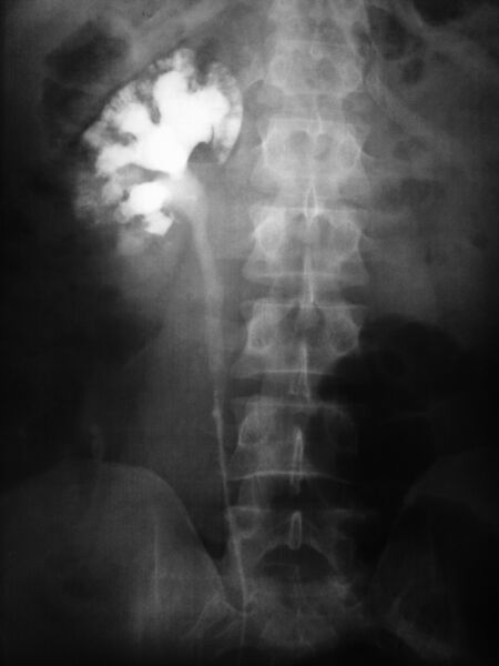
- Findings: Ectatic distal collecting ducts and also dilated contrast filled intrapapillary tubules within the renal medulla are seen on intravenous pyelogram. A case of Prof Saeed Rad, Tabriz, Iran.
- Published: 2nd Jan 2013
- Source: https://radiopaedia.org/cases/medullary-sponge-kidney-on-ivp
- Author: Mohammad Taghi Niknejad
- Permission: http://creativecommons.org/licenses/by-nc-sa/3.0/
Licensing:
Attribution-NonCommercial-ShareAlike 3.0 Unported (CC BY-NC-SA 3.0)
File history
Click on a date/time to view the file as it appeared at that time.
| Date/Time | Thumbnail | Dimensions | User | Comment | |
|---|---|---|---|---|---|
| current | 10:29, 24 March 2021 | | 886 × 1,181 (302 KB) | Fæ (talk | contribs) | Radiopaedia project rID:21065 (batch #22098) |
You cannot overwrite this file.
File usage
There are no pages that use this file.
.jpg&oldid=8856201)