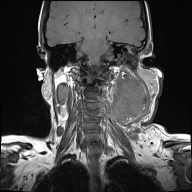
File:Nasopharyngeal carcinoma- T4,N3b (Radiopaedia 59376-67000 Coronal T1 17).jpg

From NC Commons
Jump to navigation Jump to search

Nasopharyngeal_carcinoma-_T4,N3b_(Radiopaedia_59376-67000_Coronal_T1_17).jpg(384 × 384 pixels, file size: 57 KB, MIME type: image/jpeg)

Summary:

Description
  • Radiopaedia case ID: 59376
  • Image ID: 37768345
  • Image stack position: 17/51
  • Plane projection: Coronal
  • Aux modality: T1
  • Study findings: Large left nasopharyngeal mass measuring 6.6 x 3.8 cm in bulk dimensions The mass extends into the left parapharyngeal space and inferiorly to the level of the base of the tongue. The mass extends into the left posterior nare as well as the left pterygomaxillary fissure. Altered signal in the left masticator space with signal change in the medial pterygoid muscle and abnormal enhancement in the masticator space. Fluid in the left mastoid air cells. 1.9 cm right retropharyngeal node. Bilateral cervical adenopathy.  Matted nodes along the left cervical chain (levels II, III and 1b) with bulk dimensions of 9.8 x 6.7 cm. The largest on the right is 2.3 cm at level II. Bilateral supraclavicular nodes, the largest on the left 3.1 cm and on the right a solitary 1.3 cm node. Several left intraparotid lymph nodes, the largest 4.2 cm. Fluid in the left carotid canal.  No intracranial extension.
  • Modality: MRI
  • System: Head & Neck
  • Findings: 4.7 cm mildly enhancing soft tissue mass centered on the left fossa of Rosenmüller. The mass extends anteriorly into the left posterior nares invading the left inferior turbinate. Bony erosion of the medial wall of the left maxillary antrum.  Soft tissue widening the left pterygomaxillary fissure. The fat plane between the mass and medial pterygoid muscle is obliterated suggesting masticator space extension. 1 cm satellite nodule (10 mm) along the medial wall of the left nasal cavity. No bony erosion of the base of the skull. Multiple enlarged lymph nodes at left level Ib, II, III, IV and V. Multiple left intraparotid nodes most of which have internal necrosis. The largest measures 3 cm in the left parotid gland. Multiple left supraclavicular nodes. Multiple enlarged lymph nodes at right level II, the largest one measuring 1.4 cm. Mucosal thickening of the left maxillary antrum, sphenoid and middle group of ethmoid sinuses. Right parotid, submandibular and thyroid glands are unremarkable.
Date Published: 17th Apr 2018
Source https://radiopaedia.org/cases/nasopharyngeal-carcinoma-t4n3b
Author Ian Bickle
Permission
(Permission-reusing-text)
http://creativecommons.org/licenses/by-nc-sa/3.0/

Licensing:

Attribution-NonCommercial-ShareAlike 3.0 Unported (CC BY-NC-SA 3.0)

File history

Click on a date/time to view the file as it appeared at that time.

Date/TimeThumbnailDimensionsUserComment
current23:48, 2 August 2021Thumbnail for version as of 23:48, 2 August 2021384 × 384 (57 KB) (talk | contribs)Radiopaedia project rID:59376 (thread B) (batch #25071-17 A17)