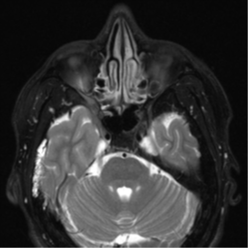
File:Nasopharyngeal carcinoma with pterygopalatine fossa involvement (Radiopaedia 33102-34134 Axial T2 22).png
Jump to navigation
Jump to search
Nasopharyngeal_carcinoma_with_pterygopalatine_fossa_involvement_(Radiopaedia_33102-34134_Axial_T2_22).png (512 × 512 pixels, file size: 220 KB, MIME type: image/png)
Summary:
| Description |
|
| Date | Published: 1st Jan 2015 |
| Source | https://radiopaedia.org/cases/nasopharyngeal-carcinoma-with-pterygopalatine-fossa-involvement |
| Author | Paul Simkin |
| Permission (Permission-reusing-text) |
http://creativecommons.org/licenses/by-nc-sa/3.0/ |
Licensing:
Attribution-NonCommercial-ShareAlike 3.0 Unported (CC BY-NC-SA 3.0)
File history
Click on a date/time to view the file as it appeared at that time.
| Date/Time | Thumbnail | Dimensions | User | Comment | |
|---|---|---|---|---|---|
| current | 05:22, 3 August 2021 |  | 512 × 512 (220 KB) | Fæ (talk | contribs) | Radiopaedia project rID:33102 (thread B) (batch #25074-22 A22) |
You cannot overwrite this file.
File usage
There are no pages that use this file.
