File:Nasopharyngeal carcinoma with skull base invasion (Radiopaedia 53415-59485 Coronal T1 39).jpg

From NC Commons
Jump to navigation Jump to search

Nasopharyngeal_carcinoma_with_skull_base_invasion_(Radiopaedia_53415-59485_Coronal_T1_39).jpg(588 × 588 pixels, file size: 63 KB, MIME type: image/jpeg)

Summary:

Description
  • Radiopaedia case ID: 53415
  • Image ID: 30394264
  • Image stack position: 39/51
  • Plane projection: Coronal
  • Aux modality: T1
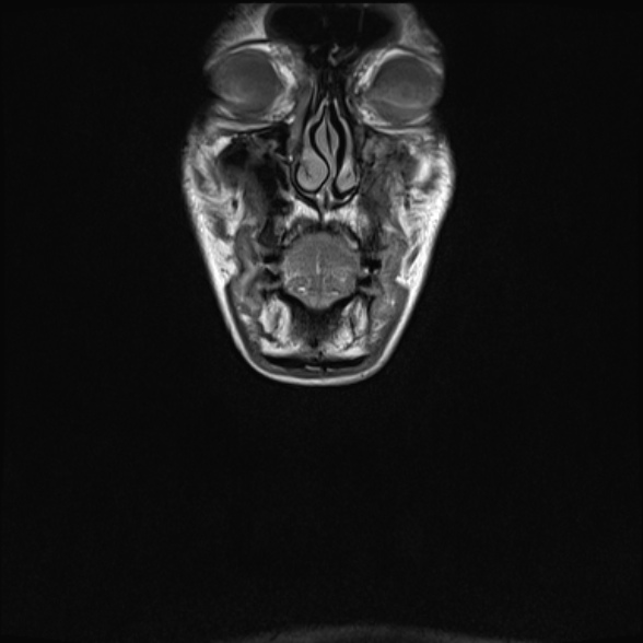
  • Study findings: Large well defined enhancing mass centered on the left fossa of Rosenmuller with bulk dimensions of 5.4 x 4.4 cm.  The mass extends inferiorly to the oropharynx and superiorly into and almost completely filling the sphenoid sinus.  On the left the tumor involves the posterior nasal canal. The mass crosses right of the midline at the level of the nasopharynx and on the left abuts 180 degrees of the internal carotid artery. A 8mm tongue of tissue extends into and widens the left sphenopalatine foramen and pterygopalatine fossa into the pterygomaxillary tissue. Pituitary fossa and floor of the anterior cranial fossa are intact.  The left medial pterygoid plate and posterior aspect of the bone nasal septum is irregular with cortical disruption. Obstructive fluid in the left maxillary sinus.  Obstructive fluid in the left mastoid due to Eustachian tube obstruction from the mass. Malignant 4.9 cm left level V nodes with some smaller adjacent sub-centimeter nodes. Left level III nodes up to 15 mm.    Left 1B up to 13 mm.
  • Modality: MRI
  • System: Head & Neck
  • Findings: Large well defined enhancing mass centered on the left fossa of Rosenmuller with bulk dimensions of 5.4 x 4.4cm.  The mass extends inferiorly on the left to the oropharynx and superiorly into and almost completely filling the sphenoid sinus.  On the left the tumor involves the posterior nasal canal. The mass crosses right of the midline at the level of the nasopharynx and on the left abuts 180 degrees of the internal carotid artery. A 8 mm tongue of tissue extends into and widens the left sphenopalatine foramen and pterygopalatine fossa into the pterygomaxillary tissue. Pituitary fossa and floor of the anterior cranial fossa are intact.  The left medial pterygoid plate and posterior aspect of the bone nasal septum is irregular with cortical disruption. Obstructive fluid in the left maxillary sinus.  Obstructive fluid in the left mastoid due to Eustachian tube obstruction from the mass. Malignant 4.9 cm left level V nodes with some smaller adjacent sub-centimeter nodes. Left level III nodes up to 15 mm.    Left 1B up to 13 mm.
Date Published: 1st Jun 2017
Source https://radiopaedia.org/cases/nasopharyngeal-carcinoma-with-skull-base-invasion
Author Ian Bickle
Permission
(Permission-reusing-text)
http://creativecommons.org/licenses/by-nc-sa/3.0/

Licensing:

Attribution-NonCommercial-ShareAlike 3.0 Unported (CC BY-NC-SA 3.0)

File history

Click on a date/time to view the file as it appeared at that time.

Date/TimeThumbnailDimensionsUserComment
current08:09, 3 August 2021Thumbnail for version as of 08:09, 3 August 2021588 × 588 (63 KB) (talk | contribs)Radiopaedia project rID:53415 (thread B) (batch #25076-39 A39)