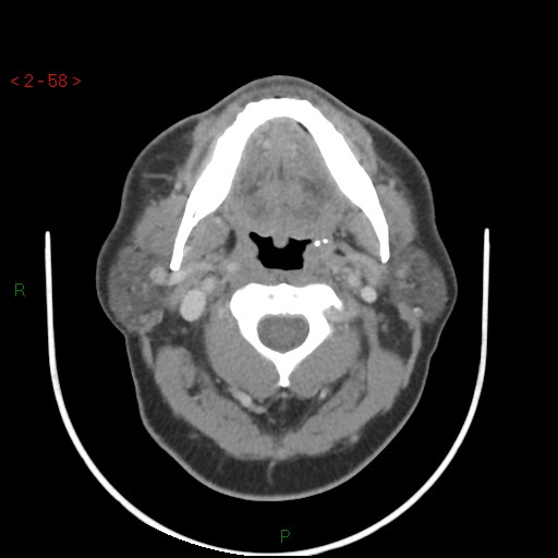
File:Nasopharyngeal lymphoma (Radiopaedia 54235-60433 A 19).jpg
Jump to navigation
Jump to search
Nasopharyngeal_lymphoma_(Radiopaedia_54235-60433_A_19).jpg (512 × 512 pixels, file size: 35 KB, MIME type: image/jpeg)
Summary:
| Description |
|
| Date | Published: 3rd Jul 2017 |
| Source | https://radiopaedia.org/cases/nasopharyngeal-lymphoma |
| Author | Safwat Mohammad Almoghazy |
| Permission (Permission-reusing-text) |
http://creativecommons.org/licenses/by-nc-sa/3.0/ |
Licensing:
Attribution-NonCommercial-ShareAlike 3.0 Unported (CC BY-NC-SA 3.0)
File history
Click on a date/time to view the file as it appeared at that time.
| Date/Time | Thumbnail | Dimensions | User | Comment | |
|---|---|---|---|---|---|
| current | 09:18, 3 August 2021 |  | 512 × 512 (35 KB) | Fæ (talk | contribs) | Radiopaedia project rID:54235 (thread B) (batch #25077-19 A19) |
You cannot overwrite this file.
File usage
There are no pages that use this file.
