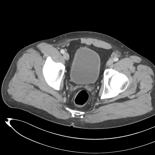
File:Necrotizing pancreatitis with acute necrotic collections (Radiopaedia 38829-41012 B 77).png
Jump to navigation
Jump to search
Necrotizing_pancreatitis_with_acute_necrotic_collections_(Radiopaedia_38829-41012_B_77).png (512 × 512 pixels, file size: 150 KB, MIME type: image/png)
Summary:
| Description |
|
| Date | Published: 6th Aug 2015 |
| Source | https://radiopaedia.org/cases/necrotising-pancreatitis-with-acute-necrotic-collections-1 |
| Author | Henry Knipe |
| Permission (Permission-reusing-text) |
http://creativecommons.org/licenses/by-nc-sa/3.0/ |
Licensing:
Attribution-NonCommercial-ShareAlike 3.0 Unported (CC BY-NC-SA 3.0)
File history
Click on a date/time to view the file as it appeared at that time.
| Date/Time | Thumbnail | Dimensions | User | Comment | |
|---|---|---|---|---|---|
| current | 23:58, 3 August 2021 |  | 512 × 512 (150 KB) | Fæ (talk | contribs) | Radiopaedia project rID:38829 (thread B) (batch #25166-135 B77) |
You cannot overwrite this file.
File usage
The following page uses this file:
