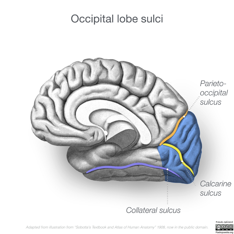
File:Neuroanatomy- medial cortex (diagrams) (Radiopaedia 47208-58969 F 1).png
Jump to navigation
Jump to search

Size of this preview: 600 × 600 pixels. Other resolutions: 240 × 240 pixels | 480 × 480 pixels | 768 × 768 pixels | 1,024 × 1,024 pixels | 2,400 × 2,400 pixels.
Original file (2,400 × 2,400 pixels, file size: 1.96 MB, MIME type: image/png)
Summary:
| Description |
|
| Date | Published: 4th Aug 2016 |
| Source | https://radiopaedia.org/cases/neuroanatomy-medial-cortex-diagrams |
| Author | Frank Gaillard |
| Permission (Permission-reusing-text) |
http://creativecommons.org/licenses/by-nc-sa/3.0/ |
Licensing:
Attribution-NonCommercial-ShareAlike 3.0 Unported (CC BY-NC-SA 3.0)
File history
Click on a date/time to view the file as it appeared at that time.
| Date/Time | Thumbnail | Dimensions | User | Comment | |
|---|---|---|---|---|---|
| current | 21:18, 5 August 2021 | | 2,400 × 2,400 (1.96 MB) | Fæ (talk | contribs) | Radiopaedia project rID:47208 (thread B) (batch #25307-23 F1) |
You cannot overwrite this file.
File usage
There are no pages that use this file.
_(Radiopaedia_47208-58969_F_1).png&oldid=907099)