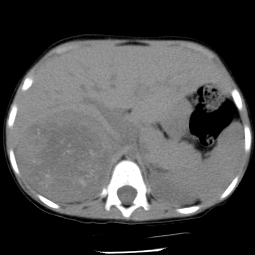
File:Neuroblastoma (Radiopaedia 7746-8572 Axial non-contrast 1).jpg
Jump to navigation
Jump to search
Neuroblastoma_(Radiopaedia_7746-8572_Axial_non-contrast_1).jpg (512 × 512 pixels, file size: 85 KB, MIME type: image/jpeg)
Summary:
| Description |
|
| Date | Published: 28th Nov 2009 |
| Source | https://radiopaedia.org/cases/neuroblastoma-3 |
| Author | Hani Makky Al Salam |
| Permission (Permission-reusing-text) |
http://creativecommons.org/licenses/by-nc-sa/3.0/ |
Licensing:
Attribution-NonCommercial-ShareAlike 3.0 Unported (CC BY-NC-SA 3.0)
File history
Click on a date/time to view the file as it appeared at that time.
| Date/Time | Thumbnail | Dimensions | User | Comment | |
|---|---|---|---|---|---|
| current | 14:31, 6 August 2021 |  | 512 × 512 (85 KB) | Fæ (talk | contribs) | Radiopaedia project rID:7746 (thread B) (batch #25330-1 A1) |
You cannot overwrite this file.
File usage
There are no pages that use this file.
