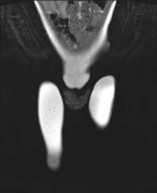
File:Neuroblastoma with bone metastases (Radiopaedia 67080-76414 Coronal T1 1).jpg
Jump to navigation
Jump to search
Neuroblastoma_with_bone_metastases_(Radiopaedia_67080-76414_Coronal_T1_1).jpg (312 × 384 pixels, file size: 14 KB, MIME type: image/jpeg)
Summary:
| Description |
|
| Date | Published: 20th Mar 2019 |
| Source | https://radiopaedia.org/cases/neuroblastoma-with-bone-metastases |
| Author | Jane McEniery |
| Permission (Permission-reusing-text) |
http://creativecommons.org/licenses/by-nc-sa/3.0/ |
Licensing:
Attribution-NonCommercial-ShareAlike 3.0 Unported (CC BY-NC-SA 3.0)
File history
Click on a date/time to view the file as it appeared at that time.
| Date/Time | Thumbnail | Dimensions | User | Comment | |
|---|---|---|---|---|---|
| current | 16:28, 6 August 2021 |  | 312 × 384 (14 KB) | Fæ (talk | contribs) | Radiopaedia project rID:67080 (thread B) (batch #25339-41 C1) |
You cannot overwrite this file.
File usage
There are no pages that use this file.
