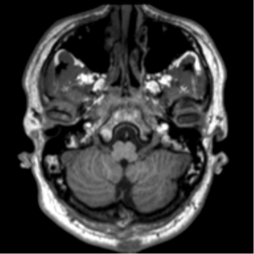
File:Neurofibromatosis type 2 (Radiopaedia 44936-48838 Axial T1 17).png

From NC Commons
Jump to navigation Jump to search

Neurofibromatosis_type_2_(Radiopaedia_44936-48838_Axial_T1_17).png(512 × 512 pixels, file size: 209 KB, MIME type: image/png)

Summary:

Description
  • Radiopaedia case ID: 44936
  • Image ID: 22171337
  • Image stack position: 17/151
  • Plane projection: Axial
  • Aux modality: T1
  • Study findings: Bilateral acoustic neuromas are noted expanding the internal acoustic meati and extending into the cerebellopontine angles in keeping with neurofibromatosis type 2. There is also an enhancing presumed schwannoma within the horizontal portion of the right facial canal in keeping with a facial nerve schwannoma. Bilateral trigeminal schwannomas are noted, on the right side this extends slightly through the foramen ovale. Further schwannomas are demonstrated related to the cranial nerves. Notably there are small bilateral oculomotor nerve schwannomas as well as an enhancing focus in the prepontine cistern on the right likely an abducens nerve schwannoma. Enhancing lesions are also noted along the course of the lower cranial nerves extending towards the jugular foramina in keeping with further lesions. Enhancement is noted centered on the central canal of the visualized upper cord, likely an ependymoma. A meningioma is noted predominantly inferior to but also extending around and superior to the left tentorium cerebelli with associated dural thickening. It compresses the quadrigeminal plate cistern, fourth ventricle and distorts the midbrain / cerebral aqueduct but without hydrocephalus. Further small meningioma is noted overlying the right frontal lobe. Bony thickening is noted in the left frontal bone near the coronal suture but no definite associated meningioma identified in this region.
  • Modality: MRI
  • System: Spine
  • Findings: Within the cervical spine there are innumerable, bilateral extramedullary, intradural masses, many of which extend through the intervertebral foramina, extending into the perivertebral muscles. These are characterized by intermediate low T1, intermediate - heterogeneous but largely high T2 signal and homogenous contrast enhancement, consistent with schwannomas. Further 3.3 cm rounded lesion of similar signal characteristic lies at the left base of the tongue, also demonstrates homogenous contrast enhancement (greater than the thyroid tissue), and likely represents a large schwannoma rather than complicated ectopic thyroid tissue. This markedly narrows the oropharynx, which is slit-like and measures 5mm at its maximal dimension. ENT assessment is suggested. Multiple other masses of similar signal characteristics are seen within the carotid vessels, and deep to both sternocleidomastoid muscles. Within the thoracolumbar spine, innumerable extramedullary, intradural schwannomas are identified, the largest of which extends through the right T3/4 intervertebral foramen measuring 3.6 x 2.9 (w x AP) cm. This demonstrates internal low T1 and high T2 signal without contrast enhancement, consistent with cystic degeneration. Within the lumbar spine, the intrathecal space is moderately narrowed at L4. Smaller schwannomas are seen within the right subcutaneous tissues, one adjacent to the right rib and many within the lumbar plexus. Conclusion: Innumerable schwannomas in keeping with neurofibromatosis type 2.
Date Published: 12th May 2016
Source https://radiopaedia.org/cases/neurofibromatosis-type-2-18
Author Frank Gaillard
Permission
(Permission-reusing-text)
http://creativecommons.org/licenses/by-nc-sa/3.0/

Licensing:

Attribution-NonCommercial-ShareAlike 3.0 Unported (CC BY-NC-SA 3.0)

File history

Click on a date/time to view the file as it appeared at that time.

Date/TimeThumbnailDimensionsUserComment
current18:18, 8 August 2021Thumbnail for version as of 18:18, 8 August 2021512 × 512 (209 KB) (talk | contribs)Radiopaedia project rID:44936 (thread B) (batch #25457-63 C17)

Metadata