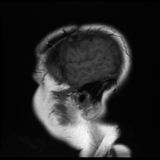
File:Neurofibromatosis type 2 (Radiopaedia 45229-49247 Sagittal T1 19).png
Jump to navigation
Jump to search
Neurofibromatosis_type_2_(Radiopaedia_45229-49247_Sagittal_T1_19).png (512 × 512 pixels, file size: 176 KB, MIME type: image/png)
Summary:
| Description |
|
| Date | Published: 19th May 2016 |
| Source | https://radiopaedia.org/cases/neurofibromatosis-type-2-6 |
| Author | Bruno Di Muzio |
| Permission (Permission-reusing-text) |
http://creativecommons.org/licenses/by-nc-sa/3.0/ |
Licensing:
Attribution-NonCommercial-ShareAlike 3.0 Unported (CC BY-NC-SA 3.0)
File history
Click on a date/time to view the file as it appeared at that time.
| Date/Time | Thumbnail | Dimensions | User | Comment | |
|---|---|---|---|---|---|
| current | 10:56, 8 August 2021 |  | 512 × 512 (176 KB) | Fæ (talk | contribs) | Radiopaedia project rID:45229 (thread B) (batch #25450-43 B19) |
You cannot overwrite this file.
File usage
There are no pages that use this file.
