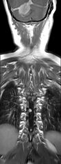
File:Neurofibromatosis type 2 (Radiopaedia 66211-75403 Coronal T1 C+ 10).jpg

From NC Commons
Jump to navigation Jump to search

Original file(512 × 1,376 pixels, file size: 238 KB, MIME type: image/jpeg)

Summary:

Description
  • Radiopaedia case ID: 66211
  • Image ID: 46419819
  • Image stack position: 10/12
  • Plane projection: Coronal
  • Aux modality: T1 C+
  • Study findings: Intramedullary lesion of the cervical spinal cord at C2 level, of low signal intensity on T1WI and high signal intensity on T2WI with no peripheral enhancement. Adjacent edema of the spinal cord is noted (above and below the lesion). Multiple enhancing intradural extramedullary lesions of various sizes along the spinal cord (largest at C2, T5-T6, and T6-T7 levels) most likely representing both meningiomas and schwannomas. Innumerable enhancing nodules scattered along the cauda equina and nerve roots, typical of schwannomas.
  • Modality: MRI
  • System: Oncology
  • Findings: Bilateral CPA soft tissue masses (larger on the left) of low signal intensity on T1WI, and isosignal intensity on T2WI and FLAIR, with intense and homogeneous enhancement, extending into an enlarged IAC bilaterally. A mass effect is noted on the middle cerebellar peduncles as well as the brainstem with low position of the cerebellar tonsils (7 mm below the foramen magnum). Extra-axial broad dural-based ovoid soft tissue mass of the posterior cerebral fossa of right paramedian location. It shows an isosignal centrally and low signal peripherally on T1WI, heterogeneous low signal on FLAIR, and central low signal with peripheral high signal on T2WI and T2*, with intense and relatively homogeneous enhancement of the central component. Enhancing soft tissue mass in the right jugular foramen, indicating schwannoma of the lower cranial nerves (IX, X, XI). Bilateral enhancing masses in the trigeminal cisterns (most likely trigeminal schwannomas). Extensive meningiomatosis with dural-based enhancing soft tissue masses along the tentorium cerebelli, falx cerebri, and cerebral convexities. Bilateral sphenoid wing dysplasia with temporal encephalocele extending downward to the infratemporal spaces, medial to the lateral pterygoid muscles. Left occipital subcutaneous lesion of low signal on T1WI and high signal on T2WI with intense and homogeneous enhancement following IV contrast. Small subcutaneous fluid collection along the course of the VP shunt at the left occipital region.
Date Published: 18th Feb 2019
Source https://radiopaedia.org/cases/neurofibromatosis-type-2-10
Author Dr Ammar Haouimi
Permission
(Permission-reusing-text)
http://creativecommons.org/licenses/by-nc-sa/3.0/

Licensing:

Attribution-NonCommercial-ShareAlike 3.0 Unported (CC BY-NC-SA 3.0)

File history

Click on a date/time to view the file as it appeared at that time.

Date/TimeThumbnailDimensionsUserComment
current14:07, 8 August 2021Thumbnail for version as of 14:07, 8 August 2021512 × 1,376 (238 KB) (talk | contribs)Radiopaedia project rID:66211 (thread B) (batch #25453-22 B10)

There are no pages that use this file.