File:Neurofibromatosis type 2 (Radiopaedia 8953-9733 C 1).jpg

From NC Commons
Jump to navigation Jump to search

Neurofibromatosis_type_2_(Radiopaedia_8953-9733_C_1).jpg(512 × 512 pixels, file size: 113 KB, MIME type: image/jpeg)

Summary:

Description
  • Radiopaedia case ID: 8953
  • Image ID: 369353
  • Image stack position: 1/21
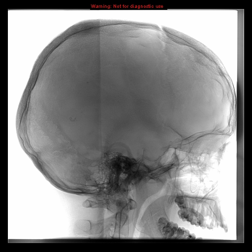
  • Study findings: The right frontal lesion prior to embolization.
  • Modality: DSA (angiography)
  • System: Oncology
  • Findings: A large right frontal extra-axial lesion is noted with homogeneous texture, homogeneous postcontrast enhancement and diffusion restriction. It has a wide dural-base with smooth scalloping of the inner table. It exerts mass effect in the form of compression of both frontal lobes, markedly on the related right side. CSF cleft is noted, confirming extra-axial location. It has prominent feeding arteries. Features are characteristic for right frontal large convexity meningioma. Right cerebellopontine cistern mass lesion with intense postcontrast enhancement, also suggestive of right CPA meningioma. Sagittal postcontrast T1WI sequence shows posterior cervical canal dural-based extra-axial lesion with homogeneous postcontrast enhancement. It is extending to the foramen magnum compressing the proximal cervical cord and cervicomedullary junction, suggestive of spinal meningioma.
Date Published: 11th Mar 2010
Source https://radiopaedia.org/cases/neurofibromatosis-type-2
Author Hani Makky Al Salam
Permission
(Permission-reusing-text)
http://creativecommons.org/licenses/by-nc-sa/3.0/

Licensing:

Attribution-NonCommercial-ShareAlike 3.0 Unported (CC BY-NC-SA 3.0)

File history

Click on a date/time to view the file as it appeared at that time.

Date/TimeThumbnailDimensionsUserComment
current15:22, 8 August 2021Thumbnail for version as of 15:22, 8 August 2021512 × 512 (113 KB) (talk | contribs)Radiopaedia project rID:8953 (thread B) (batch #25454-17 C1)

There are no pages that use this file.