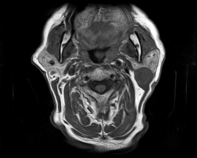
File:Non-Hodgkin lymphoma - parotid gland (Radiopaedia 71531-81890 Axial T1 11).jpg
Jump to navigation
Jump to search
Non-Hodgkin_lymphoma_-_parotid_gland_(Radiopaedia_71531-81890_Axial_T1_11).jpg (636 × 512 pixels, file size: 123 KB, MIME type: image/jpeg)
Summary:
| Description |
|
| Date | Published: 10th Oct 2019 |
| Source | https://radiopaedia.org/cases/non-hodgkin-lymphoma-parotid-gland |
| Author | Dr Ammar Haouimi |
| Permission (Permission-reusing-text) |
http://creativecommons.org/licenses/by-nc-sa/3.0/ |
Licensing:
Attribution-NonCommercial-ShareAlike 3.0 Unported (CC BY-NC-SA 3.0)
File history
Click on a date/time to view the file as it appeared at that time.
| Date/Time | Thumbnail | Dimensions | User | Comment | |
|---|---|---|---|---|---|
| current | 12:07, 11 August 2021 |  | 636 × 512 (123 KB) | Fæ (talk | contribs) | Radiopaedia project rID:71531 (thread B) (batch #25637-11 A11) |
You cannot overwrite this file.
File usage
There are no pages that use this file.
