File:Non-Hodgkin lymphoma involving seminal vesicles with development of interstitial pneumonitis during Rituximab therapy (Radiopaedia 32703-33761 ax CT Fus PET 46).jpg

From NC Commons
Jump to navigation Jump to search

Original file(1,052 × 536 pixels, file size: 58 KB, MIME type: image/jpeg)

Summary:

Description
  • Radiopaedia case ID: 32703
  • Image ID: 9629809
  • Image stack position: 46/68
  • Plane projection: ax CT Fus PET
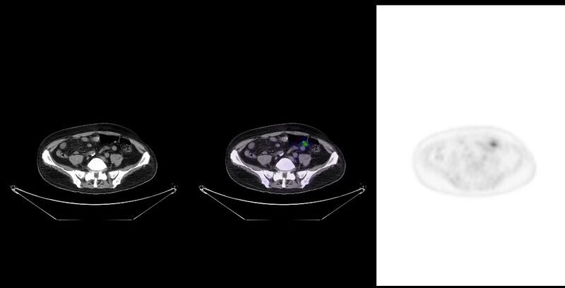
  • Study findings: All previous lesions without FDG-uptake (Deauville 1). Interval progression of ground glass opacities of posterior predominance, interspersed by normal lung tissue. Interval development of traction bronchiectasis and bronchiolectasis in affected lung. Small sub pleural cystic changes resembling early honeycombing are also noted. No overt centrilobular nodules on this low dose CT. Lung changes are moderately FDG-avid. FDG-uptake in accessory respiration muscles (scalene, and intercostal) as well as in diaphragmatic crus is also noted, consistent with dyspnea. Indwelling urinal catheter, right-sided central venous line and physiological FDG-pattern in gastrointestinal system are also noted. Conclusion Complete response. Interval progression of FDG-avid ground-glass opacities with development of traction bronchiectasis and sub pleural changes resembling hypersensitivity pneumonitis (subacute stage). Although pulmonary infection cannot be totally ruled out, findings are worrisome for Rituximab induced interstitial lung disease.
  • Modality: Nuclear medicine
  • System: Haematology
  • Findings: Enlarged and vividly FDG-enhancing seminal vesicles and appendix. FDG-avid infra- and supradiaphragmal lymphadenopathy is also noted, as well as predominantly nodular pulmonary involvement. Bilateral mild pleural effusion. No evidence of hepatic, splenic or bone marrow involvement. Incidental note is made of a gallstone in infundibulum, duodenal diverticulum, simple hepatic and renal cysts, a sub-cm lesion in the left hepatic lobe, mostly resembling hemangioma, and an umbilical hernia. Conclusion The overall imaging appearances with hyper metabolic involvement of nodal and extra nodal sites and the homogenously contrast-enhancing, but neither invasive nor vascular lesions are highly suggestive of malignant lymphoma.
Date Published: 19th Dec 2014
Source https://radiopaedia.org/cases/non-hodgkin-lymphoma-involving-seminal-vesicles-with-development-of-interstitial-pneumonitis-during-rituximab-therapy-1
Author René Pfleger
Permission
(Permission-reusing-text)
http://creativecommons.org/licenses/by-nc-sa/3.0/

Licensing:

Attribution-NonCommercial-ShareAlike 3.0 Unported (CC BY-NC-SA 3.0)

File history

Click on a date/time to view the file as it appeared at that time.

Date/TimeThumbnailDimensionsUserComment
current08:25, 11 August 2021Thumbnail for version as of 08:25, 11 August 20211,052 × 536 (58 KB) (talk | contribs)Radiopaedia project rID:32703 (thread B) (batch #25631-58 B46)