File:Non-seminomatous germ cell tumor (Radiopaedia 16649-16352 Coronal T1 C+ 8).png
Jump to navigation
Jump to search
No higher resolution available.
Non-seminomatous_germ_cell_tumor_(Radiopaedia_16649-16352_Coronal_T1_C+_8).png (512 × 512 pixels, file size: 142 KB, MIME type: image/png)
Summary:
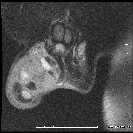
| Description |
|
| Date | Published: 7th Feb 2012 |
| Source | https://radiopaedia.org/cases/non-seminomatous-germ-cell-tumour |
| Author | Praveen Jha |
| Permission (Permission-reusing-text) |
http://creativecommons.org/licenses/by-nc-sa/3.0/ |
Licensing:
Attribution-NonCommercial-ShareAlike 3.0 Unported (CC BY-NC-SA 3.0)
File history
Click on a date/time to view the file as it appeared at that time.
| Date/Time | Thumbnail | Dimensions | User | Comment | |
|---|---|---|---|---|---|
| current | 17:59, 12 August 2021 | | 512 × 512 (142 KB) | Fæ (talk | contribs) | Radiopaedia project rID:16649 (thread B) (batch #25719-34 E8) |
You cannot overwrite this file.
File usage
There are no pages that use this file.
.png&oldid=992172)