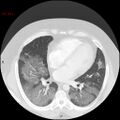
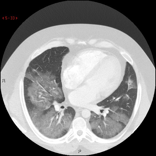
File:Non-specific interstitial pneumonitis (Radiopaedia 27044-27222 Axial lung window 24).jpg
Jump to navigation
Jump to search
Non-specific_interstitial_pneumonitis_(Radiopaedia_27044-27222_Axial_lung_window_24).jpg (512 × 512 pixels, file size: 51 KB, MIME type: image/jpeg)
Summary:
| Description |
|
| Date | Published: 18th Jan 2014 |
| Source | https://radiopaedia.org/cases/non-specific-interstitial-pneumonitis-1 |
| Author | David Preston |
| Permission (Permission-reusing-text) |
http://creativecommons.org/licenses/by-nc-sa/3.0/ |
Licensing:
Attribution-NonCommercial-ShareAlike 3.0 Unported (CC BY-NC-SA 3.0)
File history
Click on a date/time to view the file as it appeared at that time.
| Date/Time | Thumbnail | Dimensions | User | Comment | |
|---|---|---|---|---|---|
| current | 03:07, 13 August 2021 |  | 512 × 512 (51 KB) | Fæ (talk | contribs) | Radiopaedia project rID:27044 (thread B) (batch #25737-24 A24) |
You cannot overwrite this file.
File usage
The following page uses this file:
