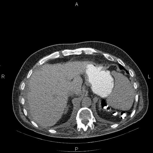
File:Non Hodgkin lymphoma in a patient with ankylosing spondylitis (Radiopaedia 84323-99624 Axial non-contrast 30).jpg

From NC Commons
Jump to navigation Jump to search

Summary:

Description
  • Radiopaedia case ID: 84323
  • Image ID: 53908752
  • Image stack position: 30/68
  • Plane projection: Axial
  • Aux modality: non-contrast
  • Study description: Control CT scan post chemotherapy.
  • Study findings: Pleural effusions present bilaterally, right more than the left side. There are also several atelectatic bands scattered bilaterally.The right kidney is relatively small than left kidney and shows relatively parenchymal atrophic changes. A few non-enhanced simple cortical cysts are seen at both kidneys, with maximum diameters of 35mm. Several stones are seen at right kidney less than 23mm causing mild hydronephrosis. Additionally, several stones are also noted at left side less than 15mm. Lymphadenopathy with SAD of 15mm is present at upper paracaval region.The prostate gland is enlarged.Degenerative changes as osteophytosis are seen at the cervicothoracolumbar spine. Additionally, diffuse ankylosis of whole spine is evident with bilateral sacroiliac joint narrowing. With respect to prior CT scan the process is relatively subsided.
  • Modality: CT
  • System: Haematology
  • Findings: Bilateral pleural effusions are present, left more than the right side, accompanied by left lower lobe partial collapse. There are no intrapulmonary nodules.Multiple lymphadenopathies are seen at mediastinum and both hilar regions with maximum SAD of 20mm. Multiple para-aortocaval, peripancreatic, celiac and mesenteric lymphadenopathies are seen. The largest one measures 57×37mm. Additionally, several lymphadenopathies are also seen at para-iliac and inguinal regions. The largest one measured 39×30mm. The spleen is enlarged and its cephalocaudal height measured 156mm.A few cortical cysts are seen at both kidneys less than 33mm. Several stones are noted at right kidney with a maximum diameter of 20mm, accompanied by mild hydronephrosis. Two 6mm and 3mm stones are also observed at left kidney without hydronephrosis. A small amount of free fluid is present. Diffuse ankylosis of thoracolumbar spine is present with bilateral sacroiliac joints narrowing.
Date Published: 27th Nov 2020
Source https://radiopaedia.org/cases/non-hodgkin-lymphoma-in-a-patient-with-ankylosing-spondylitis-1
Author Mohammad Taghi Niknejad
Permission
(Permission-reusing-text)
http://creativecommons.org/licenses/by-nc-sa/3.0/

Licensing:

Attribution-NonCommercial-ShareAlike 3.0 Unported (CC BY-NC-SA 3.0)

File history

Click on a date/time to view the file as it appeared at that time.

Date/TimeThumbnailDimensionsUserComment
current02:01, 11 August 2021Thumbnail for version as of 02:01, 11 August 2021512 × 512 (40 KB) (talk | contribs)Radiopaedia project rID:84323 (thread B) (batch #25627-30 A30)