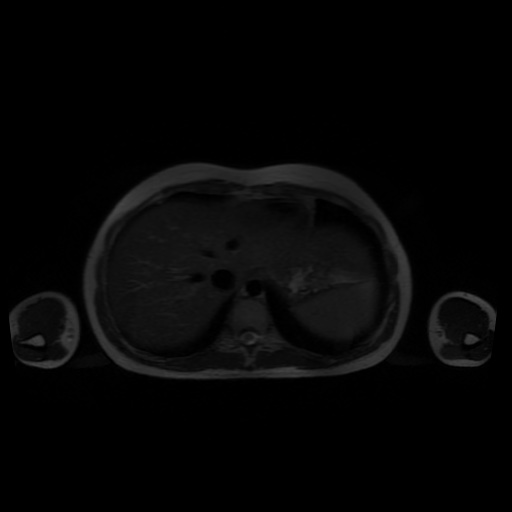
File:Normal MRI abdomen in pregnancy (Radiopaedia 88001-104541 Axial T2 6).jpg
Jump to navigation
Jump to search
Normal_MRI_abdomen_in_pregnancy_(Radiopaedia_88001-104541_Axial_T2_6).jpg (512 × 512 pixels, file size: 20 KB, MIME type: image/jpeg)
Summary:
| Description |
|
| Date | Published: 23rd Mar 2021 |
| Source | https://radiopaedia.org/cases/normal-mri-abdomen-in-pregnancy |
| Author | Vikas Shah |
| Permission (Permission-reusing-text) |
http://creativecommons.org/licenses/by-nc-sa/3.0/ |
Licensing:
Attribution-NonCommercial-ShareAlike 3.0 Unported (CC BY-NC-SA 3.0)
File history
Click on a date/time to view the file as it appeared at that time.
| Date/Time | Thumbnail | Dimensions | User | Comment | |
|---|---|---|---|---|---|
| current | 07:27, 24 August 2021 |  | 512 × 512 (20 KB) | Fæ (talk | contribs) | Radiopaedia project rID:88001 (thread B) (batch #26372-6 A6) |
You cannot overwrite this file.
File usage
The following page uses this file:
