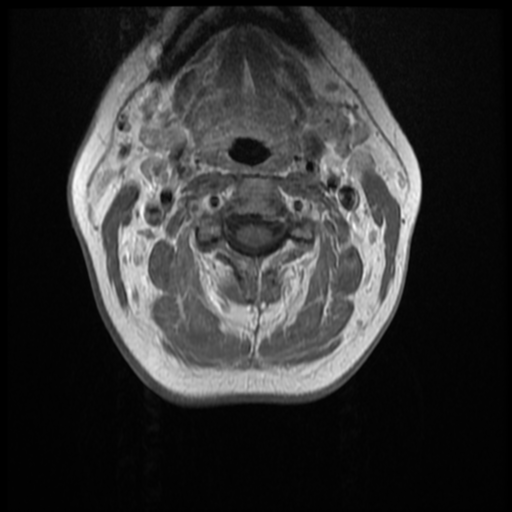
File:Normal cervical and thoracic spine MRI (Radiopaedia 35630-37156 Axial T1 C+ 24).png
Jump to navigation
Jump to search
Normal_cervical_and_thoracic_spine_MRI_(Radiopaedia_35630-37156_Axial_T1_C+_24).png (512 × 512 pixels, file size: 213 KB, MIME type: image/png)
Summary:
| Description |
|
| Date | Published: 14th Apr 2015 |
| Source | https://radiopaedia.org/cases/normal-cervical-and-thoracic-spine-mri |
| Author | Frank Gaillard |
| Permission (Permission-reusing-text) |
http://creativecommons.org/licenses/by-nc-sa/3.0/ |
Licensing:
Attribution-NonCommercial-ShareAlike 3.0 Unported (CC BY-NC-SA 3.0)
File history
Click on a date/time to view the file as it appeared at that time.
| Date/Time | Thumbnail | Dimensions | User | Comment | |
|---|---|---|---|---|---|
| current | 00:51, 20 August 2021 |  | 512 × 512 (213 KB) | Fæ (talk | contribs) | Radiopaedia project rID:35630 (thread B) (batch #25973-128 F24) |
You cannot overwrite this file.
File usage
There are no pages that use this file.
