File:Normal fetal biometry - second trimester (Radiopaedia 26450-26581 A 1).jpg

From NC Commons
Jump to navigation Jump to search

Normal_fetal_biometry_-_second_trimester_(Radiopaedia_26450-26581_A_1).jpg(669 × 303 pixels, file size: 73 KB, MIME type: image/jpeg)

Summary:

Description
  • Radiopaedia case ID: 26450
  • Image ID: 5338998
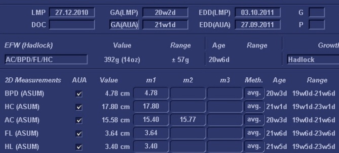
  • Study description: Biometry table
  • Study findings: The above measurements generate an ultrasound gestational age of 21w1d +/- 7d. This is compatible with the menstural gestational age of 20w2d (ie permissible 6 day discrepancy).
  • Modality: Ultrasound
  • System: Obstetrics
  • Findings: Biparietal diameter (BPD) is measured from outer cortex of the near parietal bone to the inner cortex of the far parietal bone on a symmetric image, at the level of the thalami. Cerebellar hemispheres should not be seen in this plane. The biparietal plane is near-parallel to the ultrasound beam. Head circumference (HC) is measured in this case by an elliptical tool on the same imaging plane. Abdominal circumference (AC) is measured at the level of liver, preferentially at the confluence of the right and left portal veins and with stomach on view, from outer margin of skin. Femur length (FL) measurement should be obtained with the femur perpendicular or near perpendicular to the ultrasound beam. Only the diaphysis is measured.
Date Published: 22nd Dec 2013
Source https://radiopaedia.org/cases/normal-fetal-biometry-second-trimester-1
Author Alexandra Stanislavsky
Permission
(Permission-reusing-text)
http://creativecommons.org/licenses/by-nc-sa/3.0/

Licensing:

Attribution-NonCommercial-ShareAlike 3.0 Unported (CC BY-NC-SA 3.0)

File history

Click on a date/time to view the file as it appeared at that time.

Date/TimeThumbnailDimensionsUserComment
current22:21, 22 August 2021Thumbnail for version as of 22:21, 22 August 2021669 × 303 (73 KB) (talk | contribs)Radiopaedia project rID:26450 (thread B) (batch #26187-1 A1)

There are no pages that use this file.
Metadata