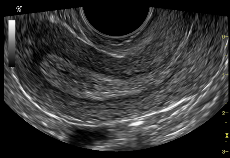
File:Normal pelvic ultrasound - transvaginal (Radiopaedia 31750-32684 B 1).png
Jump to navigation
Jump to search

Size of this preview: 800 × 551 pixels. Other resolutions: 320 × 221 pixels | 640 × 441 pixels | 1,024 × 706 pixels | 1,396 × 962 pixels.
Original file (1,396 × 962 pixels, file size: 752 KB, MIME type: image/png)
Summary:
| Description |
|
| Date | Published: 25th Oct 2014 |
| Source | https://radiopaedia.org/cases/normal-pelvic-ultrasound-transvaginal |
| Author | Bruno Di Muzio |
| Permission (Permission-reusing-text) |
http://creativecommons.org/licenses/by-nc-sa/3.0/ |
Licensing:
Attribution-NonCommercial-ShareAlike 3.0 Unported (CC BY-NC-SA 3.0)
File history
Click on a date/time to view the file as it appeared at that time.
| Date/Time | Thumbnail | Dimensions | User | Comment | |
|---|---|---|---|---|---|
| current | 19:08, 26 August 2021 | | 1,396 × 962 (752 KB) | Fæ (talk | contribs) | Radiopaedia project rID:31750 (thread B) (batch #26487-2 B1) |
You cannot overwrite this file.
File usage
There are no pages that use this file.
.png&oldid=1142682)