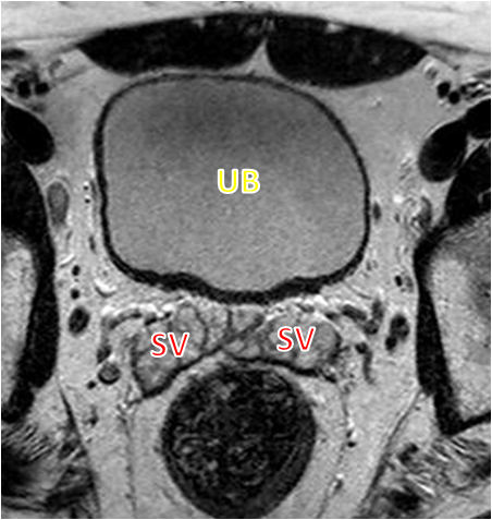
File:Normal prostate (MRI) (Radiopaedia 29986-30536 A 1).png
Jump to navigation
Jump to search
No higher resolution available.
Normal_prostate_(MRI)_(Radiopaedia_29986-30536_A_1).png (452 × 477 pixels, file size: 231 KB, MIME type: image/png)
Summary:
| Description |
|
| Date | Published: 8th Aug 2014 |
| Source | https://radiopaedia.org/cases/normal-prostate-mri-1 |
| Author | Dalia Ibrahim |
| Permission (Permission-reusing-text) |
http://creativecommons.org/licenses/by-nc-sa/3.0/ |
Licensing:
Attribution-NonCommercial-ShareAlike 3.0 Unported (CC BY-NC-SA 3.0)
File history
Click on a date/time to view the file as it appeared at that time.
| Date/Time | Thumbnail | Dimensions | User | Comment | |
|---|---|---|---|---|---|
| current | 12:00, 27 August 2021 | | 452 × 477 (231 KB) | Fæ (talk | contribs) | Radiopaedia project rID:29986 (thread B) (batch #26568-1 A1) |
You cannot overwrite this file.
File usage
The following file is a duplicate of this file (more details):
_(Radiopaedia_29986-30536_A_1).png)
_(Radiopaedia_29986-30536_B_1).png)
There are no pages that use this file.
_(Radiopaedia_29986-30536_A_1).png&oldid=1155118)